A Case Report with Plasmablastic Lymphoma of the Jejunum
- Affiliations
-
- 1Department of Internal Medicine, University of Kyunghee College of Medicine, Seoul, Korea. dramc@hanmail.net
- 2Department of Pathology, University of Kyunghee College of Medicine, Seoul, Korea.
- KMID: 1778052
- DOI: http://doi.org/10.3346/jkms.2010.25.3.496
Abstract
- Plasmablastic lymphoma (PBL) is a recently identified entity that is considered to be a type of diffuse large B-cell lymphoma with a unique immunophenotype and a predilection for the oral cavity of patients with the human immunodeficiency virus (HIV). Although its clinical features may help in the differential diagnosis, an extraoral location in a patient without HIV makes it more difficult to suspect clinically. This case report is the first to describe a patient with PBL originating from the jejunum in a 60-yr-old, HIV-seronegative man. Computed tomography of the face, chest and abdomen showed about a 9.4x9.0 cm mass of the proximal jejunum, multiple masses in the musculoskeletal soft tissue, and multiple lymphadenopathies. The histological examinations demonstrated a large cell lymphoma with plasmablastic differentiation. The neoplastic cells were diffusely positive for MUM1, epithelial membrane antigen and lambda light chains, and focally positive for CD79a; but negative for CD3, CD20, CD30, CD34, CD45RO, CD56, CD99, and CD117. The proliferation index by Ki-67 immunohistochemistry was approximately 70%. These findings were compatible with the diagnosis of PBL. The findings in this case suggest that PBL should be included in the differential diagnosis of a small bowel mass even in a HIV-negative patient.
Keyword
MeSH Terms
Figure
-

Fig. 1 Endoscopic finding. The conventional esophagogastroduodenoscopy showed a friable ulcerofungating mass over 10 cm segment.

Fig. 2 Radiologic images of a jejunal mass. (A) Abdominal CT shows a 9.4×9.0 cm enhancing lobulated mass (arrow) originating from the proximal jejunum. (B) Small bowel series shows a growing exophytic mass (arrows) with central ulcers in the proximal jejunum. The adjacent bowel loops were displaced; however, the other small bowel loops were normal. A B
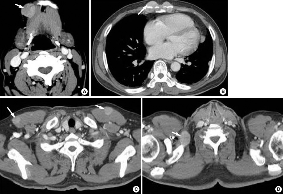
Fig. 3 CT imaging showing multiple areas of lymphoma infiltration. Arrows indicate lymphoma lesions. (A) About a 2.3 cm ovoid mass in the right submandibular space. (B, C) Multiple oval or round masses in the musculoskeletal soft tissue in the chest wall and pectoralis major muscle. (D) Chest CT shows a right supraclavicular lymphadenopathy.
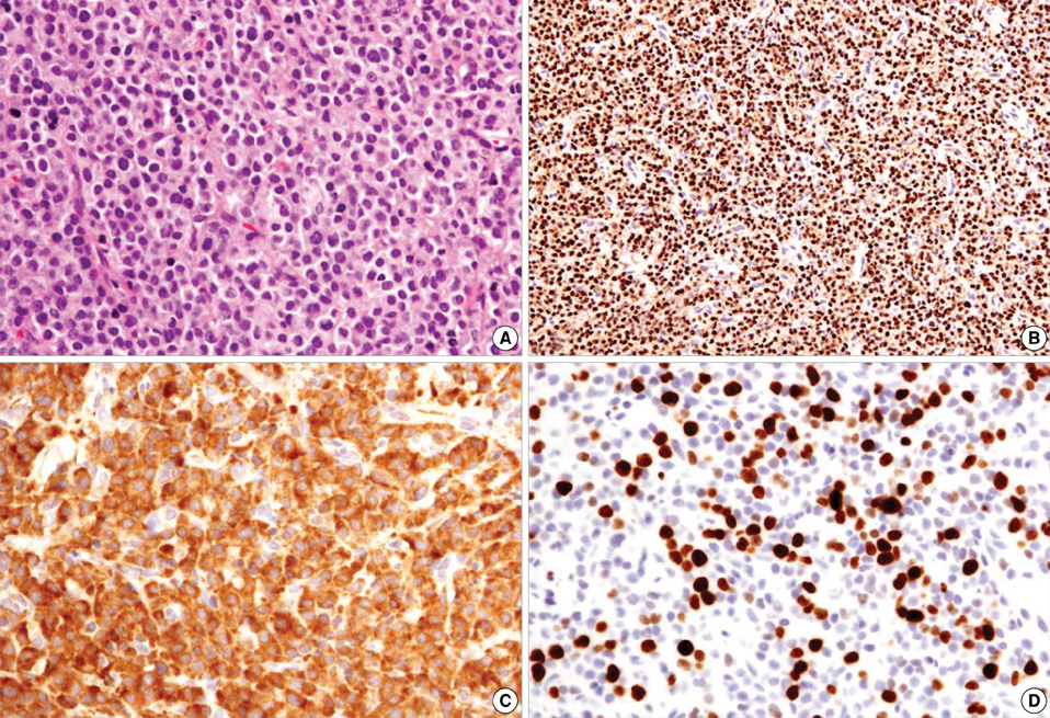
Fig. 4 Histopathology of plasmablastic lymphoma. (A) Haematoxylin and eosin section reveals a diffuse plasmablastic infiltrate with abundant cytoplasm, round nuclei, and occasionally central locating nucleoli (H&E stain, ×400). (B) The lymphoid infiltrate was positive for MUM1 (Polymer method, ×200). (C) Lambda light chain reactivity is seen in virtually all tumor cells (Polymer method, ×400). (D) The stain for Ki-67 demonstrates nuclear staining in approximately 70% of the neoplastic cells (Polymer method, ×400).
Reference
-
1. Harris NL, Jaffe ES, Diebold J, Flandrin G, Muller-Hermelink HK, Vardiman J, Lister TA, Bloomfield CD. The World Health Organization classification of neoplasms of the hematopoietic and lymphoid tissues: report of the Clinical Advisory Committee meeting-Airlie House, Virginia, November, 1997. Hematol J. 2000. 1:53–66.2. Delecluse HJ, Anagnostopoulos I, Dallenbach F, Hummel M, Marafioti T, Schneider U, Huhn D, Schmidt-Westhausen A, Reichart PA, Gross U, Stein H. Plasmablastic lymphomas of the oral cavity: a new entity associated with the human immunodeficiency virus infection. Blood. 1997. 89:1413–1420.
Article3. Flaitz CM, Nichols CM, Walling DM, Hicks MJ. Plasmablastic lymphoma: an HIV-associated entity with primary oral manifestations. Oral Oncol. 2002. 38:96–102.
Article4. Lin Y, Rodrigeus GD, Turner JF, Vasef MA. Plasmablastic lymphoma of the lung: report of a unique case and review of the literature. Arch Pathol Lab Med. 2001. 125:282–285.5. Pruneri G, Graziadei G, Ermellino L, Baldini L, Neri A, Buffa R. Plasmablastic lymphoma of the stomach: a case report. Haematologica. 1998. 83:87–89.6. Brown RS, Campbell C, Lishman SC, Spittle MF, Miller RF. Plasmablastic lymphoma: a new subcategory of human immunodeficiency virus-related non-Hodgkin's lymphoma. Clin Oncol (R Coll Radiol). 1998. 10:327–329.
Article7. Riedel DJ, Gonzalez-Cuyar LF, Zhao XF, Redfield RR, Gilliam BL. Plasmablastic lymphoma of the oral cavity: a rapidly progressive lymphoma associated with HIV infection. Lancet Infect Dis. 2008. 8:261–267.
Article8. Colomo L, Loong F, Rives S, Pittaluga S, Martínez A, López-Guillermo A, Ojanguren J, Romagosa V, Jaffe ES, Campo E. Diffuse large B-cell lymphomas with plasmablastic differentiation represent a heterogeneous group of disease entities. Am J Surg Pathol. 2004. 28:736–747.
Article9. Falini B, Fizzotti M, Pucciarini A, Bigerna B, Marafioti T, Gambacorta M, Pacini R, Alunni C, Natali-Tanci L, Ugolini B, Sebastiani C, Cattoretti G, Pileri S, Dalla-Favera R, Stein H. A monoclonal antibody (MUM1p) detects expression of the MUM1/IRF4 protein in a subset of germinal center B cells, plasma cells, and activated T cells. Blood. 2000. 95:2084–2092.
Article
- Full Text Links
-
- Actions
-
Cited
- CITED
-
- Close
- Share
-
- Similar articles
-
- Plasmablastic plasma cell myeloma mimicking plasmablastic lymphoma
- A case of plasmablastic lymphoma in the nasal cavity in a human immunodeficiency virus-negative patient
- Plasmablastic Lymphoma in a Human Immunodeficiency Virus-negative Patient: A Case Report and Review of the Literature
- Plasmablastic Lymphoma in the Anal Canal
- Concurrent intestinal plasmablastic lymphoma and diffuse large B-cell lymphoma with a clonal relationship: a case report and literature review
