A Scoring System for Prediction of Lateral Neck Node Metastasis from Papillary Thyroid Cancer
- Affiliations
-
- 1Thyroid Cancer Center, Department of Surgery, Gangnam Severance Hospital, Yonsei University College of Medicine, Seoul, Korea. surghsc@yuhs.ac
- 2Department of Biostatistics, Yonsei University College of Medicine, Seoul, Korea.
- KMID: 1777836
- DOI: http://doi.org/10.3346/jkms.2011.26.8.996
Abstract
- Lateral neck node metastasis is an important prognostic factor in thyroid carcinoma. We developed a scoring system for use in prediction of lateral neck node metastasis from papillary thyroid cancer. In this study, 161 consecutive patients were included in the training data set. This scoring system, named the Yonsei Estimated Value (YEV) for lymph node metastasis in papillary thyroid cancer, was developed on the basis of results from multivariate logistic regression analysis of preoperative clinical and radiologic data. Sixty eight consecutive patients were included for testing of the validity of the scoring system. The equation for prediction of lateral neck node metastasis was follows: YEV (Yonsei Estimated Value) = 1/(1+X) X = Exp (5.333-[0.902 x sex]+[0.036 x age]-[1.020 x tumor size]-[0.177 x lymph node size]-[0.032 x lymph node density]) When the YEV was 0.3 or more, the probability of lateral neck node metastasis was 79.0%, with sensitivity of 76.3%, specificity of 69.8%, positive predictive value of 56.7%, and negative predictive value of 85.1% in the training set. When fine needle aspiration biopsy for suspicious lateral neck nodes is not possible, or the results are inadequate, our scoring system for prediction of lateral neck node metastasis can be helpful in optimization of the surgical extent for each patient.
Keyword
MeSH Terms
Figure
-

Fig. 1 Schematic figure of the X axis and Y axis. The lymph node is targeted according to the location in the axial plane of CT, with the Y axis defined as the distance from the sternum to the target lymph node, and the X axis is as the distance from the upper Y-axis point to the target lymph node.

Fig. 2 CT findings for lateral neck lymph nodes are CT density after contrast injection (Hounsfield Unit, HU) of the lateral neck lymph node. (A) proven nonmetastatic node, with 108 HU. (B) proven metastatic node, with 164 HU.
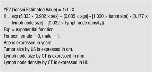
Fig. 3 Scoring system. CT, computed tomography; US, ultrasound; HU, Hounsfield units.
Cited by 1 articles
-
Overcoming the Limitations of Fine Needle Aspiration Biopsy: Detection of Lateral Neck Node Metastasis in Papillary Thyroid Carcinoma
Hak Hoon Jun, Seok Mo Kim, Bup Woo Kim, Yong Sang Lee, Hang-Seok Chang, Cheong Soo Park
Yonsei Med J. 2015;56(1):182-188. doi: 10.3349/ymj.2015.56.1.182.
Reference
-
1. Harwood J, Clark OH, Dunphy JE. Significance of lymph node metastasis in differentiated thyroid cancer. Am J Surg. 1978. 136:107–112.2. Hughes CJ, Shaha AR, Shah JP, Loree TR. Impact of lymph node metastasis in differentiated carcinoma of the thyroid: a matched-pair analysis. Head Neck. 1996. 18:127–132.3. King AD, Tse GM, Ahuja AT, Yuen EH, Vlantis AC, To EW, van Hasselt AC. Necrosis in metastatic neck nodes: diagnostic accuracy of CT, MR imaging, and US. Radiology. 2004. 230:720–726.4. Sarvanan K, Bapuraj JR, Sharma SC, Radotra BD, Khandelwal N, Suri S. Computed tomography and ultrasonographic evaluation of metastatic cervical lymph nodes with surgicoclinicopathologic correlation. J Laryngol Otol. 2002. 116:194–199.5. Jeong HS, Baek CH, Son YI, Choi JY, Kim HJ, Ko YH, Chung JH, Baek HJ. Integrated 18F-FDG PET/CT for the initial evaluation of cervical node level of patients with papillary thyroid carcinoma: comparison with ultrasound and contrast-enhanced CT. Clin Endocrinol (Oxf). 2006. 65:402–407.6. Lee SK, Choi JH, Lim HI, Kim WW, Kim SM, Choe JH, Lee JE, Shin JH, Choi JY, Kim JH, Kim JS, Nam SJ, Yang JH. Sentinel lymph node biopsy in papillary thyroid cancer: comparison study of blue dye method and combined radioisotope and blue dye method in papillary thyroid cancer. Eur J Surg Oncol. 2009. 35:974–979.7. Mirallié E, Sagan C, Hamy A, Paineau J, Kahn X, Le Néel JC, Auget JL, Murat A, Joubert M, Le Bodic MF, Visset J. Predictive factors for node involvement in papillary thyroid carcinoma. Univariate and multivariate analyses. Eur J Cancer. 1999. 35:420–423.8. Lim CY, Sohn EJ, Lee J, Yun JS, Nam KH, Chang HS, Chung WY, Park CS. The significant predicting factors influencing lateral neck node metastasis in papillary thyroid carcinoma. J Korean Surg Soc. 2006. 71:326–330.9. Mancuso AA, Harnsberg HR, Muraki AS, Stevens MA. Computed tomography of cervical and retropharyngeal lymph nodes: normal anatomy and, variants of normal, and application in stging head and neck cancer. Part II: pathology. Radiology. 1983. 148:715–723.10. Som PM, Brandwein M, Lidov M, Lawson W, Biller HF. The varied presentations of papillary thyroid carcinoma cervical nodal disease: CT and MR findings. AJNR Am J Neuroradiol. 1994. 15:1123–1128.11. Ito Y, Tomoda C, Uruno T, Takamura Y, Miya A, Kobayashi K, Matsuzuka F, Kuma K, Miyauchi A. Ultrasonographically and anatomopathologically detectable node metastases in the lateral compartment as indicators of worse relapse-free survival in patients with papillary thyroid carcinoma. World J Surg. 2005. 29:917–920.12. Kim EH, Park JS, Son KR, Kim JH, Jeon SJ, Na DG. Preoperative diagnosis of cervical metastatic lymph nodes in papillary thyroid carcinoma: comparison of ultrasound, computed tomography, and combined ultrasound with computed tomography. Thyroid. 2008. 18:411–418.13. Kang TW, Shin JH, Han BK, Ko EY, Kang SS, Hahn SY, Kim JS, Oh YL. Preoperative ultrasound-guided tattooing localization of recurrences after thyroidectomy: safety and effectiveness. Ann Surg Oncol. 2009. 16:1655–1659.14. Kim MJ, Kim EK, Kim BM, Kwak JY, Lee EJ, Park CS, Cheong WY, Nam KH. Thyroglobulin measurement in fine-needle aspirate washouts: the criteria for neck node dissection for patients with thyroid cancer. Clin Endocrinol (Oxf). 2009. 70:145–151.
- Full Text Links
-
- Actions
-
Cited
- CITED
-
- Close
- Share
-
- Similar articles
-
- Chyle Leakage After Right Selective Lymph Node Dissection in a Patient With Papillary Thyroid Cancer: A Case Report
- Retropharyngeal Lymph Node Metastasis from Thyroid Papillary Carcinoma with Airway Obstruction
- Regional Lymph Node Metastasis in Papillary Thyroid Cancer
- The Pattern of Cervical Lymph Node Metastases in Papillary Thyroid Cancer
- Ultrasonographic Features of Metastatic Lymph Nodes in Papillary Thyroid Microcarcinomas and Macrocarcinomas
