Diagnostic Importance of 3D CT Images in Klippel-Feil Syndrome with Multiple Skeletal Anomalies: A Case Report
- Affiliations
-
- 1Kahramanmaras Sutcu Imam University, Radiology Dept., Turkey. myuksel@ksu.edu.tr
- 2Kahramanmaras Sutcu Imam University, Pediatrics Dept., Turkey.
- 3Kahramanmaras Sutcu Imam University, Neuroradiology Dept., Turkey.
- KMID: 1777286
- DOI: http://doi.org/10.3348/kjr.2005.6.4.278
Abstract
- We present here the case of a 12-year-old boy who had Klippel-Feil syndrome with renal, cardiac and multiple skeletal anomalies, and we show the relevent three-dimensional computed tomography images. Our patient had a triple renal pelvis, mitral valve prolapsus, multiple cervical vertebrae fusions, cervical ribs, hypoplasia of the right thumb, spina bifida of L5, lumbalization at the right side of S1 and a sacral curved defect. In this study, we discuss the atypical clinical features and the diagnostic value of three-dimensional CT for evaluating the skeletal anomalies of the Klippel-Feil syndrome cases.
MeSH Terms
Figure
-
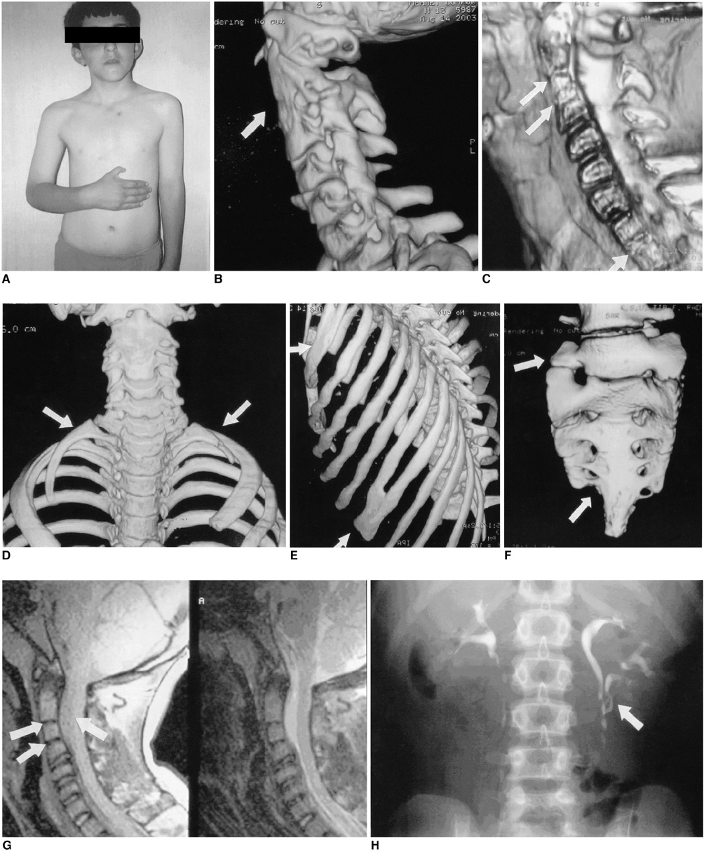
Fig. 1 A. General appearance of the patient with hypoplasia of the right thumb, and asymmetry of the chest wall and shoulders. B. Volume rendering three-dimensional CT lateral cervical image showing the fusion (arrow) of the vertebral bodies and the posterior elements of C2-C4 with the wasp-waist sign. C. Volume rendering three-dimensional CT sagittal cervical image showing the fusion (arrows) of C2-4 and T1-2 (arrow). D. Volume rendering three-dimensional CT chest image showing bilateral cervical ribs (arrows) at the level of C7. E. Volume rendering three-dimensional CT chest image showing the rib fusion at the right side, T1-2 and T8-9 levels (arrows). F. Volume rendering three-dimensional CT lumbar image showing the sacral defect with the absence of the S5 foramina and the lumbalization (arrows) at the right side of S1. G. T1 and T2 weighted sagittal images showing cervical fusion with enlargement of spinal canal and the dural sac ectasia (arrows). H. Intravenous urography showing a triple renal pelvis (arrow) in the left kidney.
Reference
-
1. Nguyen VD, Tyrrel R. Klippel-Feil syndrome: Patterns of bony fusion and wasp-waist sign. Skeletal Radiol. 1993. 22:519–523.2. Thomsen MN, Schneider U, Weber M, Johannisson R, Niethard FU. Scoliosis and congenital anomalies associated with Klippel-Feil syndrome types I-III. Spine. 1997. 22:396–401.3. Vaidyanathan S, Hughes PL, Soni BM, Singh G, Sett P. Klippel-Feil syndrome - the risk of cervical spinal cord injury: a case report. BMC Fam Pract. 2002. 3:6.4. Pizzutillo PD, Woods M, Nicholson L, MacEwen GD. Risk factors in Klippel-Feil syndrome. Spine. 1994. 19:2110–2116.5. Dahnert W. Radiology Review Manual. 1993. 2nd ed. Maryland: Williams & Wilkins;130.6. Moore WB, Matthews TJ, Rabinowitz R. Genitourinary anomalies associated with Klippel-Feil syndrome. J Bone Joint Surg Am. 1975. 57:355–357.7. Masuda H, Arikawa K, Yuda T, Taira A. Total anomalous pulmonary venous connection associated with Klippel-Feil syndrome: a case report] (abstract). Kyobu Geka. 1991. 44:417–420.8. Algom M, Schlesinger Z. Prolapse of the mitral valve in Klippel-Feil syndrome. Chest. 1981. 79:127–128.9. Schaefer-Prokop C, von Smekal U, van der Molen AJ. Prokop M, Galanski M. Musculoskeletal system. Spiral and multislice computed tomography of the body. 2003. Stuttgart, NewYork: Thieme;929–997.10. Schlosser RJ, Faust RA, Phillips CD, Gross CW. Three-dimensional computed tomography of congenital nasal anomalies. Int J Pediatr Otorhinolaryngol. 2002. 65:125–131.
- Full Text Links
-
- Actions
-
Cited
- CITED
-
- Close
- Share
-
- Similar articles
-
- A Case of Klippel-Feil Syndrome with Recurrent Hypoglycemia
- Traumatic Hemiparesis Associated with Type III Klippel-Feil Syndrome
- Sprengel Deformity with Bilateral Huge Omovertebra
- Endotracheal Intubation Using McGrath Videolaryngoscope in Klippel-Feil Syndrome
- Progressive Quadriparesis following a Minor Trauma in a Patient with Klippel-Feil Syndrome: Case Report
