Korean J Radiol.
2009 Aug;10(4):416-419. 10.3348/kjr.2009.10.4.416.
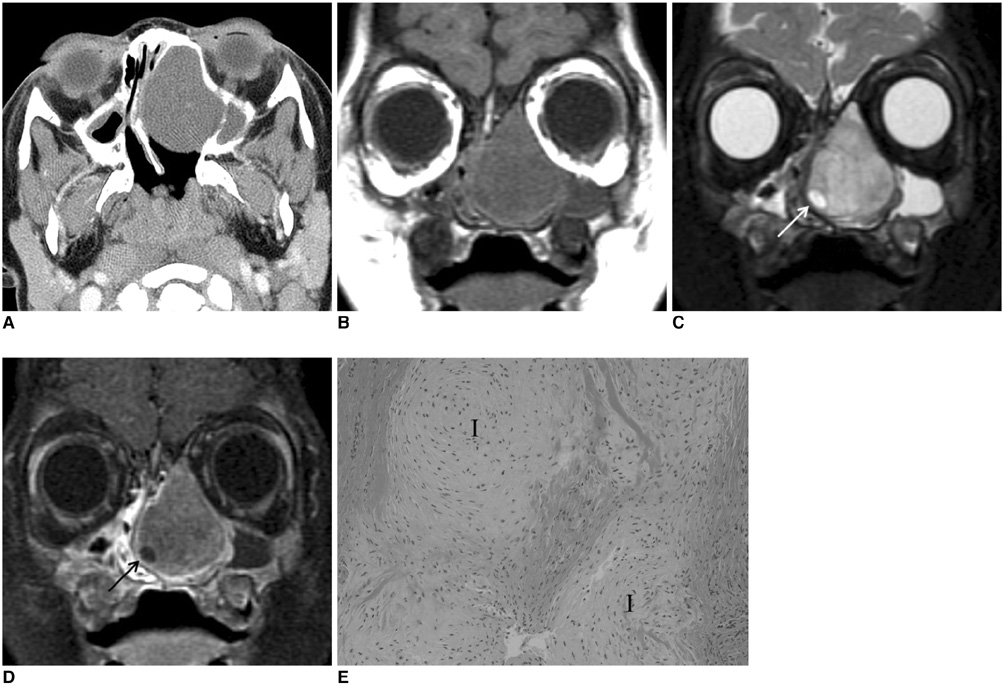
Nasal Chondromesenchymal Hamartoma: CT and MR Imaging Findings
- Affiliations
-
- 1Department of Radiology, Samsung Medical Center, Sungkyunkwan University School of Medicine, Seoul 135-710, Korea. hyungkim@skku.edu
- 2Department of Pathology, Samsung Medical Center, Sungkyunkwan University School of Medicine, Seoul 135-710, Korea.
- 3Department of Otorhinolaryngology-Head and Neck Surgery, Samsung Medical Center, Sungkyunkwan University School of Medicine, Seoul 135-710, Korea.
- KMID: 1777273
- DOI: http://doi.org/10.3348/kjr.2009.10.4.416
Abstract
- We report CT and MR imaging findings for a case of nasal chondromesenchymal hamartoma occurring in a 19-month-old boy. A nasal chondromesenchymal hamartoma is a rare benign pediatric hamartoma that can simulate malignancy. Although rare, knowledge of this entity is essential to avoid potentially harmful therapies.
MeSH Terms
Figure
Cited by 1 articles
-
Three Cases of Nasal Chondromesenchymal Hamartoma Occurred in Sinonasal Tract
Yeonjoo Choi, Yong Ju Jang, Kyung-Ja Cho, Yoo-Sam Chung
Korean J Otorhinolaryngol-Head Neck Surg. 2019;62(11):651-656. doi: 10.3342/kjorl-hns.2018.00815.
Reference
-
1. McDermott MB, Ponder TB, Dehner LP. Nasal chondromesenchymal hamartoma: an upper respiratory tract analogue of the chest wall mesenchymal hamartoma. Am J Surg Pathol. 1998. 22:425–433.2. Alrawi M, McDermott M, Orr D, Russell J. Nasal chondromesenchymal hamartoma presenting in an adolescent. Int J Pediatr Otorhinolaryngol. 2003. 67:669–672.3. Norman ES, Bergman S, Trupiano JK. Nasal chondromesenchymal hamartoma: report of a case and review of the literature. Pediatr Dev Pathol. 2004. 7:517–520.4. Ozolek JA, Carrau R, Barnes EL, Hunt JL. Nasal chondromesenchymal hamartoma in older children and adults: series and immunohistochemical analysis. Arch Pathol Lab Med. 2005. 129:1444–1450.5. Johnson C, Nagaraj U, Esguerra J, Wasdahl D, Wurzbach D. Nasal chondromesenchymal hamartoma: radiographic and histopathologic analysis of a rare pediatric tumor. Pediatr Radiol. 2007. 37:101–104.6. Kim DW, Low W, Billman G, Wickersham J, Kearns D. Chondroid hamartoma presenting as a neonatal nasal mass. Int J Pediatr Otorhinolaryngol. 1999. 47:253–259.7. Hsueh C, Hsueh S, Gonzalez-Crussi F, Lee T, Su J. Nasal chondromesenchymal hamartoma in children: report of 2 cases with review of the literature. Arch Pathol Lab Med. 2001. 125:400–403.8. Kim B, Park SH, Min HS, Rhee JS, Wang KC. Nasal chondromesenchymal hamartoma of infancy clinically mimicking meningoencephalocele. Pediatr Neurosurg. 2004. 40:136–140.9. Silkiss RZ, Mudvari SS, Shetlar D. Ophthalmologic presentation of nasal chondromesenchymal hamartoma in an infant. Ophthal Plast Reconstr Surg. 2007. 23:243–244.10. Finitsis S, Giavroglou C, Potsi S, Constantinidis I, Mpaltatzidis A, Rachovitsas D, et al. Nasal chondromesenchymal hamartoma in a child. Cardiovasc Intervent Radiol. 2009. 32:593–597.
- Full Text Links
- Actions
-
Cited
- CITED
-
- Close
- Share
- Similar articles
-
- Nasal Chondromesenchymal Hamartoma: A case report
- Three Cases of Nasal Chondromesenchymal Hamartoma Occurred in Sinonasal Tract
- A Rare Case of Recurrent Myoid Hamartoma Mimicking Malignancy: Imaging Appearances
- MR Findings of Hamartoma of the Breast: A Report of Two Cases
- Hypothalamic hamartoma associated with precocious puberty: case report


