Reliability of RTVue, Visante, and Slit-Lamp Adapted Ultrasonic Pachymetry for Central Corneal Thickness Measurement
- Affiliations
-
- 1Department of Ophthalmology, Yonsei University College of Medicine, Seoul, Korea. eungkkim@yuhs.ac
- 2Department of Ophthalmology, CHA Bundang Medical Center, CHA University, Seongnam, Korea.
- 3Corneal Dystrophy Research Institute, Yonsei University College of Medicine, Seoul, Korea.
- 4Department of Surgical Specialties Ophthalmology Clinic, University Hospital G. Martino, University of Messina, Messina, Italy.
- 5Severance Biomedical Science Institute, Brain Korea 21 Project for Medical Science, Yonsei University College of Medicine, Seoul, Korea.
- KMID: 1777001
- DOI: http://doi.org/10.3349/ymj.2012.53.3.634
Abstract
- PURPOSE
To evaluate reliability of Fourier-domain optical coherence tomography (OCT) (RTVue), time-domain OCT (Visante), and slit-lamp adapted ultrasonic pachymetry (SL-US) in the measurement of central corneal thickness (CCT).
MATERIALS AND METHODS
Thirty healthy volunteers visited our clinic 3 times and fifty eyes were measured by one physician. RTVue and Visante were randomly performed, and then SL-US, in which the ultrasound probe was inserted into the Goldmann tonometry mount, was done. During the second visit, each measurement was repeated 3 times. Measurements on the second visit were averaged, and agreement among the instruments was investigated with Bland-Altman plots.
RESULTS
RTVue showed smaller repeatability coefficient than Visante and SL-US (4.7, 8.3, and 7.7 microm, respectively). Intersession reproducibility of RTVue and Visante was worse than their repeatability. CCT of RTVue was estimated to be maximally different by 11.8 microm from CCT of Visante and 8.8 microm from CCT of SL-US. The repeatability coefficient of SL-US was 7.7 microm and its reproducibility was similar to the repeatability.
CONCLUSION
CCT measured by RTVue showed good reliability and generally agreed with Visante and SL-US. SL-US was as reliable as triple touching conventional US even with a single touch on the cornea.
MeSH Terms
Figure
-
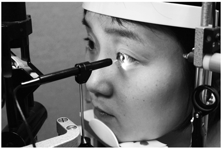
Fig. 1 Demonstration of slit-lamp adapted ultrasonic pachymetry. The ultrasound probe is inserted into the Goldmann tonometry mount, and under joystick control, it can be softly and precisely used to touch the surface of the cornea.
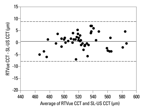
Fig. 2 Bland-Altman plot of the difference in the measurement of central corneal thickness between RTVue and slit-lamp adapted ultrasonic pachymetry (SL-US). Mean difference of 0.5±4.3 (corrected SD) µm with limits of agreement between - 7.8 to 8.8 µm. For each subject, three consecutive measurements (repeatability data) with each instrument were averaged, and the difference was calculated. CCT, central corneal thickness; SD, standard deviation.
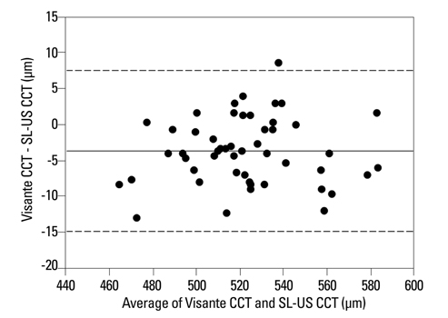
Fig. 3 Bland-Altman plot of the difference in the measurement of central corneal thickness between Visante and slit-lamp adapted ultrasonic pachymetry (SL-US). Mean difference of - 3.6±5.7 (corrected SD) µm with limits of agreement between - 14.9 to 7.6 µm. For each subject, three consecutive measurements (repeatability data) with each instrument were averaged, and the difference was calculated. CCT, central corneal thickness; SD, standard deviation.
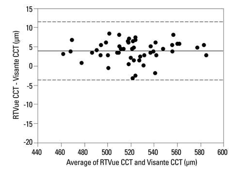
Fig. 4 Bland-Altman plot of the difference in the measurement of central corneal thickness between RTVue and Visante. Mean difference of 4.1±3.9 (corrected SD) µm with limits of agreement between - 3.5 to 11.8 µm. For each subject, three consecutive measurements (repeatability data) with each instrument were averaged, and the difference was calculated. CCT, central corneal thickness; SD, standard deviation.
Cited by 1 articles
-
Comparison of Preoperative and Postoperative Ocular Biometry in Eyes with Phakic Intraocular Lens Implantations
Joo Youn Shin, Jae Bum Lee, Kyoung Yul Seo, Eung Kweon Kim, Tae-im Kim
Yonsei Med J. 2013;54(5):1259-1265. doi: 10.3349/ymj.2013.54.5.1259.
Reference
-
1. Doughty MJ, Zaman ML. Human corneal thickness and its impact on intraocular pressure measures: a review and meta-analysis approach. Surv Ophthalmol. 2000. 44:367–408.
Article2. Higginbotham EJ, Gordon MO, Beiser JA, Drake MV, Bennett GR, Wilson MR, et al. The Ocular Hypertension Treatment Study: topical medication delays or prevents primary open-angle glaucoma in African American individuals. Arch Ophthalmol. 2004. 122:813–820.3. Chihara E. Assessment of true intraocular pressure: the gap between theory and practical data. Surv Ophthalmol. 2008. 53:203–218.
Article4. Leem HS, Lee KJ, Shin KC. Central corneal thickness and corneal endothelial cell changes caused by contact lens use in diabetic patients. Yonsei Med J. 2011. 52:322–325.
Article5. Ashwin PT, McDonnell PJ. Collagen cross-linkage: a comprehensive review and directions for future research. Br J Ophthalmol. 2010. 94:965–970.
Article6. Nam SM, Lee HK, Kim EK, Seo KY. Comparison of corneal thickness after the instillation of topical anesthetics: proparacaine versus oxybuprocaine. Cornea. 2006. 25:51–54.
Article7. Li H, Leung CK, Wong L, Cheung CY, Pang CP, Weinreb RN, et al. Comparative study of central corneal thickness measurement with slit-lamp optical coherence tomography and visante optical coherence tomography. Ophthalmology. 2008. 115:796–801.
Article8. Mohamed S, Lee GK, Rao SK, Wong AL, Cheng AC, Li EY, et al. Repeatability and reproducibility of pachymetric mapping with Visante anterior segment-optical coherence tomography. Invest Ophthalmol Vis Sci. 2007. 48:5499–5504.
Article9. Huang D, Swanson EA, Lin CP, Schuman JS, Stinson WG, Chang W, et al. Optical coherence tomography. Science. 1991. 254:1178–1181.
Article10. Kaluzny BJ, Kałuzny JJ, Szkulmowska A, Gorczyńska I, Szkulmowski M, Bajraszewski T, et al. Spectral optical coherence tomography: a novel technique for cornea imaging. Cornea. 2006. 25:960–965.11. Muscat S, McKay N, Parks S, Kemp E, Keating D. Repeatability and reproducibility of corneal thickness measurements by optical coherence tomography. Invest Ophthalmol Vis Sci. 2002. 43:1791–1795.12. Reliablity (statistics). Wikipedia. accessed 2011 August 1. Avaliable at: http://en.wikipedia.org/wiki/Reliability_(statistics).13. International vocabulary of metrology - Basic and general concepts and associated terms (VIM). JCGM 200:2008. accessed 2011 August 1. Avaliable at: http://www.iso.org/sites/JCGM/VIM/JCGM_200e.html.14. Bland JM, Altman DG. Measurement error. BMJ. 1996. 313:744.15. Bland JM, Altman DG. Statistical methods for assessing agreement between two methods of clinical measurement. Lancet. 1986. 1:307–310.
Article16. Bland JM, Altman DG. Agreement between methods of measurement with multiple observations per individual. J Biopharm Stat. 2007. 17:571–582.
Article17. Bland JM. What is the standard error of the within-subject standard deviation, sw? accessed 2011 August 1. Avaliable at: http://www-users.york.ac.uk/~mb55/meas/seofsw.htm.18. Bland JM, Altman DG. Measurement error proportional to the mean. BMJ. 1996. 313:106.19. Garson GD. Reliability Analysis. accessed 2011 August 1. Avaliable at: http://faculty.chass.ncsu.edu/garson/PA765/reliab.htm#rater.20. Realini T, Lovelace K. Measuring central corneal thickness with ultrasound pachymetry. Optom Vis Sci. 2003. 80:437–439.
Article21. Brandt JD, Beiser JA, Kass MA, Gordon MO. Central corneal thickness in the Ocular Hypertension Treatment Study (OHTS). Ophthalmology. 2001. 108:1779–1788.
Article22. Miglior S, Albe E, Guareschi M, Mandelli G, Gomarasca S, Orzalesi N. Intraobserver and interobserver reproducibility in the evaluation of ultrasonic pachymetry measurements of central corneal thickness. Br J Ophthalmol. 2004. 88:174–177.
Article23. O'Donnell C, Maldonado-Codina C. Agreement and repeatability of central thickness measurement in normal corneas using ultrasound pachymetry and the OCULUS Pentacam. Cornea. 2005. 24:920–924.24. Lackner B, Schmidinger G, Pieh S, Funovics MA, Skorpik C. Repeatability and reproducibility of central corneal thickness measurement with Pentacam, Orbscan, and ultrasound. Optom Vis Sci. 2005. 82:892–899.
Article25. Amano S, Honda N, Amano Y, Yamagami S, Miyai T, Samejima T, et al. Comparison of central corneal thickness measurements by rotating Scheimpflug camera, ultrasonic pachymetry, and scanning-slit corneal topography. Ophthalmology. 2006. 113:937–941.
Article26. Prydal JI, Artal P, Woon H, Campbell FW. Study of human precorneal tear film thickness and structure using laser interferometry. Invest Ophthalmol Vis Sci. 1992. 33:2006–2011.27. Prospero Ponce CM, Rocha KM, Smith SD, Krueger RR. Central and peripheral corneal thickness measured with optical coherence tomography, Scheimpflug imaging, and ultrasound pachymetry in normal, keratoconus-suspect, and post-laser in situ keratomileusis eyes. J Cataract Refract Surg. 2009. 35:1055–1062.
Article28. Zhao PS, Wong TY, Wong WL, Saw SM, Aung T. Comparison of central corneal thickness measurements by visante anterior segment optical coherence tomography with ultrasound pachymetry. Am J Ophthalmol. 2007. 143:1047–1049.
Article29. Chen S, Huang J, Wen D, Chen W, Huang D, Wang Q. Measurement of central corneal thickness by high-resolution Scheimpflug imaging, Fourier-domain optical coherence tomography and ultrasound pachymetry. Acta Ophthalmol. 2010. [Epub ahead of print].
Article30. Huang JY, Pekmezci M, Yaplee S, Lin S. Intra-examiner repeatability and agreement of corneal pachymetry map measurement by time-domain and Fourier-domain optical coherence tomography. Graefes Arch Clin Exp Ophthalmol. 2010. 248:1647–1656.
Article31. Nam SM, Im CY, Lee HK, Kim EK, Kim TI, Seo KY. Accuracy of RTVue optical coherence tomography, Pentacam, and ultrasonic pachymetry for the measurement of central corneal thickness. Ophthalmology. 2010. 117:2096–2103.
Article32. Prakash G, Agarwal A, Jacob S, Kumar DA, Agarwal A, Banerjee R. Comparison of fourier-domain and time-domain optical coherence tomography for assessment of corneal thickness and intersession repeatability. Am J Ophthalmol. 2009. 148:282–290.
Article
- Full Text Links
-
- Actions
-
Cited
- CITED
-
- Close
- Share
-
- Similar articles
-
- Measurement of Anterior Segment Using Visante OCT in Koreans
- The Effects of Dry Eye on the Corneal Thickness Measured by Orbscan and Ultrasonic Pachymetry
- Comparison of Corneal Thickness Measurements Between Orbscan Topography System and Ultrasonic Pachymetry before and after LASIK
- Central Corneal Thickness Measured by Four Different Methods in Normal and Post-Femtosecond Laser-Assisted LASIK Eyes
- Accuracy of Orbscan Pachymetry Measurements and Ultrasonic Pachymetry before and after LASIK with Orbscan II(R) Topography
