Digital Thermography of the Fingers and Toes in Raynaud's Phenomenon
- Affiliations
-
- 1Division of Rheumatology, Department of Internal Medicine, Inha University Hospital, Incheon, Korea. parkwon@inha.ac.kr
- 2Department of Occupational and Environmental Medicine, Inha University Hospital, Incheon, Korea.
- KMID: 1774454
- DOI: http://doi.org/10.3346/jkms.2014.29.4.502
Abstract
- The aim of this study was to determine whether skin temperature measurement by digital thermography on hands and feet is useful for diagnosis of Raynaud's phenomenon (RP). Fifty-seven patients with RP (primary RP, n = 33; secondary RP, n = 24) and 146 healthy volunteers were recruited. After acclimation to room temperature for 30 min, thermal imaging of palmar aspect of hands and dorsal aspect of feet were taken. Temperature differences between palm (center) and the coolest finger and temperature differences between foot dorsum (center) and first toe significantly differed between patients and controls. The area under curve analysis showed that temperature difference of the coolest finger (cutoff value: 2.2degrees C) differentiated RP patients from controls (sensitivity/specificity: 67/60%, respectively). Temperature differences of first toe (cutoff value: 3.11degrees C) also discriminated RP patients (sensitivity/specificity: about 73/66%, respectively). A combination of thermographic assessment of the coolest finger and first toe was highly effective in men (sensitivity/specificity : about 88/60%, respectively) while thermographic assessment of first toe was solely sufficient for women (sensitivity/specificity: about 74/68%, respectively). Thermographic assessment of the coolest finger and first toe is useful for diagnosing RP. In women, thermography of first toe is highly recommended.
MeSH Terms
Figure
-
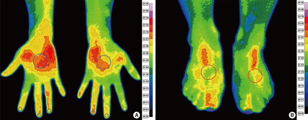
Fig. 1 Thermographic images of palmar aspect of hands (A) and dorsal aspect of feet (B). The region of interest of the distal finger/toe, palm of the hands and dorsum of the feet are drawn in circles. The color chart provides a temperature scale. First, the coolest finger was chosen among the second, third and fourth fingers and then temperature difference was determined by subtracting the temperature at the coolest finger from the temperature at the palm. Temperature difference of first toe was determined in the same manner as in the hand.
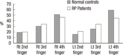
Fig. 2 The distribution of coolest finger in normal controls and RP patients. Rt/Lt denotes right/left, respectively.
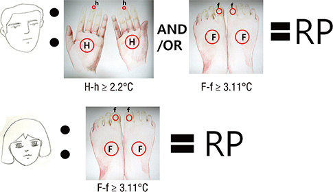
Fig. 3 The schematic summary of infrared thermography in Raynaud's phenomenon (RP). H, palm of the hand; h, coolest finger; F, dorsum of the foot; f, first toe.
Reference
-
1. Coffman JD. Raynaud's phenomenon. New York: Oxford University Press;1989.2. Jones BF. A reappraisal of the use of infrared thermal image analysis in medicine. IEEE Trans Med Imaging. 1998; 17:1019–1027.3. Block JA, Sequeira W. Raynaud's phenomenon. Lancet. 2001; 357:2042–2048.4. Herrick AL, Clark S. Quantifying digital vascular disease in patients with primary Raynaud's phenomenon and systemic sclerosis. Ann Rheum Dis. 1998; 57:70–78.5. Lambova S, Hermann W, Müller-Ladner U. Capillaroscopic pattern at the toes of systemic sclerosis patients: does it "tell" more than those of fingers? J Clin Rheumatol. 2011; 17:311–314.6. Sari-Kouzel H, Hutchinson CE, Middleton A, Webb F, Moore T, Griffin K, Herrick AL. Foot problems in patients with systemic sclerosis. Rheumatology (Oxford). 2001; 40:410–413.7. LeRoy EC, Medsger TA Jr. Raynaud's phenomenon: a proposal for classification. Clin Exp Rheumatol. 1992; 10:485–488.8. Chikura B, Moore TL, Manning JB, Vail A, Herrick AL. Sparing of the thumb in Raynaud's phenomenon. Rheumatology (Oxford). 2008; 47:219–221.9. Fleiss JL. Reliability of measurement. In : Fleiss JL, editor. The design and analysis of clinical experiments. New York: John Wiley & Sons;1986. p. 1–32.10. Coffman JD. Raynaud's phenomenon: an update. Hypertension. 1991; 17:593–602.11. La Montagna G, Baruffo A, Tirri R, Buono G, Valentini G. Foot involvement in systemic sclerosis: a longitudinal study of 100 patients. Semin Arthritis Rheum. 2002; 31:248–255.12. Anderson ME, Moore TL, Lunt M, Herrick AL. The 'distal-dorsal difference': a thermographic parameter by which to differentiate between primary and secondary Raynauds phenomenon. Rheumatology (Oxford). 2007; 46:533–538.13. Clark S, Hollis S, Campbell F, Moore T, Jayson M, Herrick A. The "distal-dorsal difference" as a possible predictor of secondary Raynaud's phenomenon. J Rheumatol. 1999; 26:1125–1128.14. Schlager O, Gschwandtner ME, Herberg K, Frohner T, Schillinger M, Koppensteiner R, Mlekusch W. Correlation of infrared thermography and skin perfusion in Raynaud patients and in healthy controls. Microvasc Res. 2010; 80:54–57.15. Heslop J, Coggon D, Acheson ED. The prevalence of intermittent digital ischaemia (Raynaud's phenomenon) in a general practice. J R Coll Gen Pract. 1983; 33:85–89.16. Gardner-Medwin JM, Macdonald IA, Taylor JY, Riley PH, Powell RJ. Seasonal differences in finger skin temperature and microvascular blood flow in healthy men and women are exaggerated in women with primary Raynaud's phenomenon. Br J Clin Pharmacol. 2001; 52:17–23.17. Pauling JD, Flower V, Shipley JA, Harris ND, McHugh NJ. Influence of the cold challenge on the discriminatory capacity of the digital distal-dorsal difference in the thermographic assessment of Raynauds phenomenon. Microvasc Res. 2011; 82:364–368.18. Do JH, Kim HY. Increased plasma endothelin-1 and abnormal nailfold capillaroscopic findings in patients with connective tissue diseases. Korean J Med. 2004; 66:275–283.
- Full Text Links
-
- Actions
-
Cited
- CITED
-
- Close
- Share
-
- Similar articles
-
- Pulsed Radiofrequency Lesioning of the Stellate Ganglion in Raynaud's Syndrome: A case report
- A Case of Raynaud's Phenomenon of both Feet in a Rock Drill Operator with Hand-arm Vibration Syndrome
- The Primary Biliary Cirrhosis Presenting with Raynaud's Disease and Digital Necrosis
- A Case of Acquired Digital Fibrokeratoma
- Digital Reconstruction with Toe Transfer
