Semi-Automatic Measurement of the Airway Dimension by Computed Tomography Using the Full-With-Half-Maximum Method: a Study of the Measurement Accuracy according to the Orientation of an Artificial Airway
- Affiliations
-
- 1Department of Radiology and Research Institute of Radiology, University of Ulsan College of Medicine, Asan Medical Center, Seoul, Korea. seojb@amc.seoul.kr
- 2Department of Industrial Engineering, Seoul National University, Seoul, Korea.
- KMID: 1758457
- DOI: http://doi.org/10.3348/kjr.2008.9.3.236
Abstract
OBJECTIVE
To develop an algorithm to measure the dimensions of an airway oriented obliquely on a volumetric CT, as well as assess the effect of the imaging parameters on the correct measurement of the airway dimension. MATERIALS AND METHODS: An airway phantom with 11 poly-acryl tubes of various lumen diameters and wall thicknesses was scanned using a 16-MDCT (multidetector CT) at various tilt angles (0, 30, 45, and 60degree). The CT images were reconstructed at various reconstruction kernels and thicknesses. The axis of each airway was determined using the 3D thinning algorithm, with images perpendicular to the axis being reconstructed. The luminal radius and wall thickness was measured by the full-width-half-maximum method. The influence of the CT parameters (the size of the airways, obliquity on the radius and wall thickness) was assessed by comparing the actual dimension of each tube with the estimated values. RESULTS: The 3D thinning algorithm correctly determined the axis of the oblique airway in all tubes (mean error: 0.91 +/- 0.82degree). A sharper reconstruction kernel, thicker image thickness and larger tilt angle of the airway axis resulted in a significant decrease of the measured wall thickness and an increase of the measured luminal radius. Use of a standard kernel and a 0.75-mm slice thickness resulted in the most accurate measurement of airway dimension, which was independent of obliquity. CONCLUSION: The airway obliquity and imaging parameters have a strong influence on the accuracy of the airway wall measurement. For the accurate measurement of airway thickness, the CT images should be reconstructed with a standard kernel and a 0.75 mm slice thickness.
Keyword
MeSH Terms
Figure
-

Fig. 1 CT images of poly-acryl airway phantom. A. CT image of phantom without tilt angle. B. CT image of phantom tilted at 45° to scan plane. These images were taken with 360-mm reconstruction field of view, B50f reconstruction kernel, and 0.75-mm slice thickness. Note tube shape distortion to ovoid at tilted orientations.

Fig. 2 Software design and representative images for measurement of oblique airway. A. Schematic work flow diagram for measurement of oblique airway. B. Medial axis estimation of artificial airway tilted at 45° to scan plane. Line represents estimated medial axis on volume rendering images. C. Original axial slice of artificial airway tilted at 45° to scan plane. D. Reconstructed image orthogonal to axis of artificial airway.

Fig. 3 Effect of tilt angle and its influence on accuracy of airway wall measurement. All images were reconstructed with 0.75-mm slice thickness, standard reconstruction kernel (B50f), and 360-mm field of view. No significant difference in accuracy of airway wall measurements was observed at four different tilt angles. Airway thicknesses are overestimated for all images if airway thickness is smaller than 1 mm.

Fig. 4 Interaction of obliquity and slice thickness in measurement of wall thickness. As image thickness increases and larger tilt angle of airway axis is used, estimated wall thickness becomes smaller than actual wall thickness. 0.75-mm wall thickness provided highest quality CT image which in turn results in most accurate measurement, independent of obliquity of airway. Images were reconstructed via standard reconstruction kernel (B50f).
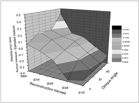
Fig. 5 Interaction of obliquity and reconstruction kernel on measurement of wall thickness. Images were reconstructed with 0.75 mm wall thickness. When sharper reconstruction kernel is used and airway is tilted to larger angle, estimated wall thickness becomes smaller than actual wall thickness. Hence, measurement of reconstructed CT images using standard kernel (B50f) results in most accurate measurement, independent of obliquity of airway.

Fig. 6 Interaction of obliquity and slice thickness on measurement of luminal radius. As image thickness increases and larger tilt angle of airway axis is used, estimated luminal radius becomes smaller than actual luminal radius. Measurement on CT image with 0.75-mm thickness results in most accurate measurement, independent of obliquity of airway. Images were reconstructed using standard reconstruction kernel (B50f).

Fig. 7 Interaction of obliquity and reconstruction kernel in accurate measurement of luminal radius. Images were reconstructed at 0.75 mm wall thickness. When sharper reconstruction kernel is used and airway is tilted to larger angle, estimated radius becomes smaller than actual luminal radius. Hence, measurement of reconstructed CT images, using standard kernel (B50f) results in most accurate measurement, independent of obliquity of airway.
Cited by 3 articles
-
Quantitative CT Imaging in Chronic Obstructive Pulmonary Disease: Review of Current Status and Future Challenges
Young Hoon Cho, Joon Beom Seo, Sang Min Lee, Sang Min Lee, Jooae Choe, Dabee Lee, Namkug Kim
J Korean Soc Radiol. 2018;78(1):1-12. doi: 10.3348/jksr.2018.78.1.1.An Engineering View on Megatrends in Radiology: Digitization to Quantitative Tools of Medicine
Namkug Kim, Jaesoon Choi, Jaeyoun Yi, Seungwook Choi, Seyoun Park, Yongjun Chang, Joon Beom Seo
Korean J Radiol. 2013;14(2):139-153. doi: 10.3348/kjr.2013.14.2.139.Prediction of Treatment Response in Patients with Chronic Obstructive Pulmonary Disease by Determination of Airway Dimensions with Baseline Computed Tomography
Hyo Jung Park, Sang Min Lee, Jooae Choe, Sang Min Lee, Namkug Kim, Jae Seung Lee, Yeon-Mok Oh, Joon Beom Seo
Korean J Radiol. 2019;20(2):304-312. doi: 10.3348/kjr.2018.0204.
Reference
-
1. Weibel ER, Taylor CR. Design and structure of human lung. Pulmonary disease and disorders. 1988. New York, NY: McGraw-Hill;11–60.2. Peter JB, Trevor TH. Prospects for new drugs for chronic obstructive pulmonary disease. Lancet. 2004. 364:985–996.3. D'Souza ND, Reinhardt JM, Hoffman EA. ASAP: interactive quantification of 2D airway geometry. SPIE Medical Imaging. 1996. 2709:180–196.4. Awadh N, Müller NL, Park CS, Abboud RT, FitzGerald JM. Airway wall thickness in patients with near fatal asthma and control groups: assessment with high resolution computed tomographic scanning. Thorax. 1998. 53:248–253.5. Brown RH, Herold CJ, Hirshman CA, Zerhouni EA, Mitzner W. In vivo measurements of airway reactivity using high-resolution computed tomography. Am Rev Respir Dis. 1991. 144:208–212.6. Brown RH, Zerhouni EA, Mitzner W. Variability in the size of individual airways over the course of one year. Am J Respir Crit Care Med. 1995. 151:1159–1164.7. McNamara AE, Müller NL, Okazawa M, Arntorp J, Wiggs BR, Paré PD. Airway narrowing in excised canine lungs measured by high-resolution computed tomography. J Appl Physiol. 1992. 73:307–316.8. Saba OI, Hoffman EA, Reinhardt JM. Maximizing quantitative accuracy of lung airway lumen and wall measures obtained from X-ray CT imaging. J Appl Physiol. 2003. 95:1063–1075.9. Kim DY, Chung SM, Park JW. Automatic navigation path generation based on two-phase adaptive region-growing algorithm for virtual angioscopy. Med Eng Phys. 2006. 28:339–347.10. Ferguson JS, McLennan G. Virtual bronchoscopy. Proc Am Thorac Soc. 2005. 2:488–491. 504–505.11. Kim N, Seo JB, Song KS, Chae EJ, Kang SH. Semi-automatic measurement of the airway dimension at computed tomography using the full-width-half-maximum method: study on the measurement accuracy according to CT parameters and size of the airway. Korean J Radiol. 2008. 9:226–235.
- Full Text Links
-
- Actions
-
Cited
- CITED
-
- Close
- Share
-
- Similar articles
-
- Semi-Automatic Measurement of the Airway Dimension by Computed Tomography Using the Full-Width-Half-Maximum Method: a Study on the Measurement Accuracy according to the CT Parameters and Size of the Airway
- Quantitative Analysis of Airway Walls Using CT Software
- Three-dimensional evaluation of the relationship between nasopharyngeal airway shape and adenoid size in children
- Anatomical Measurement of The Upper Airway Dimensions with Computed Tomography
- Airway Measurement for Airway Remodeling Defined by Post-Bronchodilator FEV1/FVC in Asthma: Investigation Using Inspiration-Expiration Computed Tomography
