J Breast Cancer.
2007 Jun;10(2):162-168. 10.4048/jbc.2007.10.2.162.
Introduction of Electrochemical Therapy (EChT) and Application of EChT to The Breast Tumor
- Affiliations
-
- 1Department of General Surgery, Konyang University Hospital, Daejeon, Korea.
- 2Department of Pathology, Konyang University Hospital, Daejeon, Korea.
- 3Department of Radiology, Konyang University Hospital, Daejeon, Korea.
- 4Department of Pharmacology, Konyang University Hospital, Daejeon, Korea.
- 5Department of Anesthesiology & Pain Medicine, Konyang University Hospital, Daejeon, Korea. gangsi@kyuh.co.kr
- 6Department of Thoracic Surgery, China-Japan Friendship Hospital, Beijing 100029, China.
- KMID: 1745063
- DOI: http://doi.org/10.4048/jbc.2007.10.2.162
Abstract
-
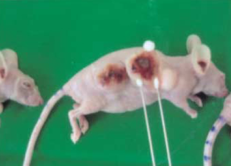
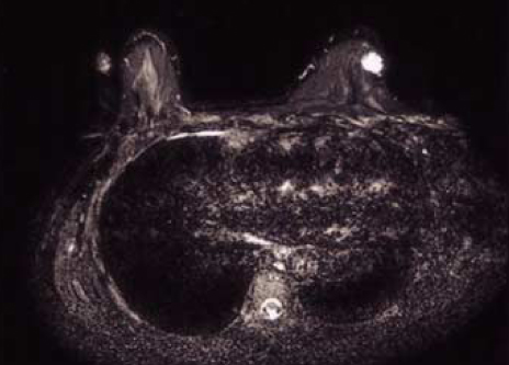
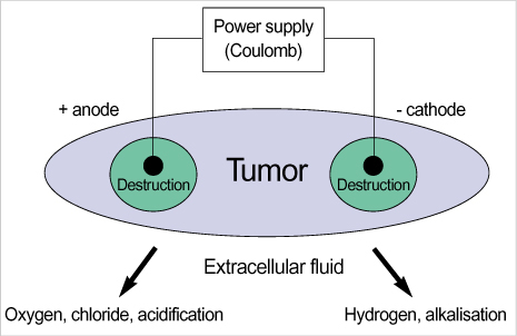
PURPOSE: To introduce the history and principle mechanism of electrochemical treatment (EChT) with animal study and report two cases successfully treated breast cancer and hemangioma by EChT.
METHODS
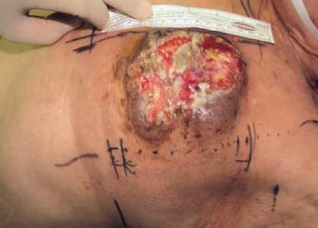
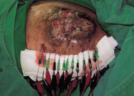
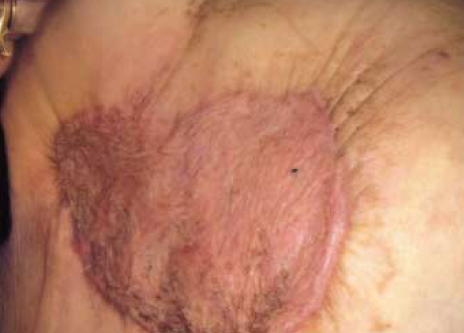
In animal study, the breast cancer tumor in nude mouse treated with EChT (100 Coulomb/cm3) were reviewed for histologic changes. In the case studies, we reported method of EChT and clinical results after EChT. Case 1: 74 yr old female with locally advanced breast cancer received 3 times EChT with 1,000 Coulomb/time, 8 Volt. Case 2: 51 yr old female with breast hemagioma received one time EChT with 80 Coulomb, 8 Volt.
RESULTS
In animal study, There were destructive change including vaculated cell fragment and extensive coagulative necrosis. Case 1 showed no local recurrence during 18 monthes after EChT. Case 2 also showed no evidence of recurrence of hemangioma.
CONCLUSION
The EChT is easy to use. It is effective, safe, less traumatic and makes patients recover quickly. This is a new and effective method to treat patients with tumours that are inoperable and can not receive chemotherapy or radiotherapy.
Keyword
MeSH Terms
Figure
Reference
-
1. Crussel G. Die Electrilytishen Heilanstalt in Moscow. Med. Zeitung Russlands. 1847. 4:2041.2. Schechter DC. Flashbacks: containment of tumors through electricity. Pacing Clin Electrophysiol. 1979. 2:100–114.
Article3. Watson BW. The treatment of tumours with direct electric current. Medical Science Research. 1991. 19:103–105.4. Nordenström BE. Survey of mechanisms in electrochemical treatment (ECT) of cancer. Eur J Surg Suppl. 1994. 574:93–109.5. Nilsson E, von Euler H, Berendson J, Thörne A, Wersäll P, Näslund I, et al. Electrochemical treatment of tumours. Bioelectrochemistry. 2000. 51:1–11.
Article6. Humphrey CE, Seal EH. Biophysical approach toward tumor regression in mice. Science. 1959. 130:388–390.
Article7. Li K, Xin Y, Gu Y, Xu B, Fan D, Ni B. Effects of direct current on dog liver: possible mechanisms for tumor electrochemical treatment. Bioelectromagnetics. 1997. 18:2–7.
Article8. Nordenström BE. Biologically closed electric circuits: clinical, experimental and theoretical evidence for an additional circulatory system. 1983. Stockholm: Nordic Medical Publications.9. Nordenström BE. Electrochemical treatment of cancer. I: Variable response to anodic and cathodic fields. Am J Clin Oncol. 1989. 12:530–536.10. Nordenström BE, Eksborg S, Beving H. Electrochemical treatment of cancer. II: Effect of electrophoretic influence on adriamycin. Am J Clin Oncol. 1990. 13:75–88.11. Xin YL. Advances in the treatment of malignant tumours by electrochemical therapy (ECT). Eur J Surg Suppl. 1994. 574:31–35.12. Samuelsson L, Jonsson L. Electrolyte destruction of lung tissue. Electrochemical aspects. Acta Radiol Diagn. 1980. 21:711–714.13. Samuelsson L, Jonsson L. Electrolytic destruction of tissue in the normal lung of the pig. Acta Radiol Diagn. 1981. 22:9–14.
Article14. Eksborg S, Nordenstrom BE, Beving H. Electrochemical treatment of cancer. III: Plasma pharmacokinetics of adriamycin after intraneoplastic administration. Am J Clin Oncol. 1990. 13:164–166.15. Yabushita H, Yoshikawa K, Hirata M, Furuya H, Hojyoh T, Fukatsu H, et al. Effects of electrochemotherapy on CaSki cells derived from a cervical squamous cell carcinoma. Gynecol Oncol. 1997. 65:297–303.
Article16. Rebersek M, Cufer T, Cemazar M, Kranjc S, Sersa G. Electroche-motherapy with cisplatin of cutaneous tumor lesions in breast cancer. Anticancer Drugs. 2004. 15:593–597.
Article17. Liu D, Xin YL, Ge B, Zhao F, Zhao H. Experimental studies on electrolytic dosage of ECT for dog's oesophageal injury and clinical effects of ECT for oesophageal anastomotic opening stenosis and oesophageal carcinoma. Eur J Surg Suppl. 1994. 574:71–72.18. Xin YL. Organisation and spread of electrochemical therapy (ECT) in China. Eur J Surg Suppl. 1994. 574:25–29.19. Xin YL, Xue F, Ge B, Zhao F, Shi B, Zhang W. Electrochemical treatment of lung cancer. Bioelectromagnetics. 1997. 18:8–13.
Article
- Full Text Links
- Actions
-
Cited
- CITED
-
- Close
- Share
- Similar articles
-
- Circulating Tumor Cells: Detection Methods and Potential Clinical Application in Breast Cancer
- The Progress and Clinical Application of Breast Cancer Organoids
- New Findings on Breast Cancer Stem Cells: A Review
- Mono- and Combination Chemotherapy for Metastatic Breast Cancer: An Increamental Step Forward
- Systemic adjuvant therapy in breast cancer

