Pineal Gland Metastasis as the Initial Presentation of Squamous Cell Lung Cancer: A Case Report
- Affiliations
-
- 1Department of Radiology, Chungnam National University Hospital, Chungnam National University School of Medicine, Daejeon, Korea. cjsong@cnu.ac.kr
- KMID: 1736473
- DOI: http://doi.org/10.3348/jksr.2014.71.3.103
Abstract
- The pineal gland is an unusual site for brain metastasis, and a solitary pineal gland metastasis is rare. A 71-year-old man presented with dizziness, gait disturbance, and memory impairment. Brain computed tomography revealed a solitary hyperdense mass with central calcification in the pineal region and obstructive hydrocephalus. Brain magnetic resonance images showed a heterogeneously enhancing mass with intratumoral calcification and microcysts. Metastatic squamous cell carcinoma was diagnosed following an endoscopic biopsy. A systemic review revealed that the primary site of the carcinoma was the lung. Although rare, metastasis should be considered in the differential diagnosis of pineal region tumors, especially in elderly patients with a pineal tumor that presents unusual imaging findings.
MeSH Terms
Figure
-
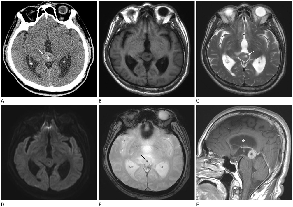
Fig. 1 A non-contrast-enhanced axial CT image shows a solitary well-defined hyperdense (50 Hounsfield units) mass (arrow, A) with central calcification in the pineal region. A well-defined lobulated mass in the pineal area shows intemediate signal intensity on axial T1 weighted images (WI) (B) and isosignal intensity on T2WI (C). There is no restricted diffusion on diffusion-weighted imaging (D). The mass contains a central dark signal intensity focus due to the engulfment of pineal calcification on gradient echo image (arrow, E), and T2 high signal intensity foci of microcysts (arrow, C). Contrast-enhanced sagittal T1WI (F) shows heterogeneous enhancement of the tumor. The pineal mass results in obstructive hydrocephalus (asterisk, F).
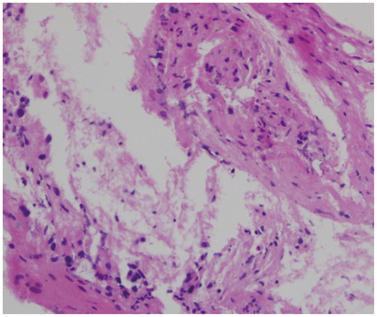
Fig. 2 Histopathologic examination (hematoxylin and eosin stain, × 200) show proliferation of atypical stratified squamous cells with keratin, which is suggestive of squamous cell carcinoma.
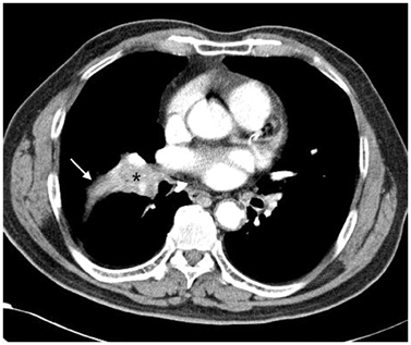
Fig. 3 A contrast-enhanced chest CT image shows a central mass (asterisk) in the right middle lobe of the lung and associated atelectasis (arrow).
Reference
-
1. Smith AB, Rushing EJ, Smirniotopoulos JG. From the archives of the AFIP: lesions of the pineal region: radiologic-pathologic correlation. Radiographics. 2010; 30:2001–2020.2. Nemoto K, Aoshiba K, Itoh M, Semba S, Tsuji T, Adachi H, et al. Isolated pineal region metastasis from lung adenocarcinoma with obstructive hydrocephalus: a case report. J Med Case Rep. 2013; 7:71.3. Vaquero J, Martínez R, Magallón R, Ramiro J. Intracranial metastases to the pineal region. Report of three cases. J Neurosurg Sci. 1991; 35:55–55.4. Oztekin O, Savas R, Ozan E, Apaydin M, Yasar O, Adibelli ZH, et al. Pineal gland metastasis of auricular squamous cell carcinoma: an unusual case and literature review. Radiol Oncol. 2009; 43:175–179.5. Samanci Y, Iplikcioglu C, Ozek E, Ozcan D, Marangozoglu B. Lung carcinoma metastasis presenting as a pineal region tumor. Neurocirugia (Astur). 2011; 22:579–582.6. Ahn JY, Chung YS, Kwon SO, Huh R, Chung SS. Isolated pineal region metastasis of small cell lung cancer. J Clin Neurosci. 2005; 12:691–693.7. Ortega P, Malamud N, Shimkin MB. Metastasis to the pineal body. AMA Arch Pathol. 1951; 52:518–528.8. Kashiwagi S, Hatano M, Yokoyama T. Metastatic small cell carcinoma to the pineal body: case report. Neurosurgery. 1989; 25:810–813.
- Full Text Links
-
- Actions
-
Cited
- CITED
-
- Close
- Share
-
- Similar articles
-
- Synchronous Primary Lung Cancer with Differrent Squamous cell Carcinoma: One Case Report
- Intrathyroidal metastasis of tonsillar squamous cell carcinoma masquerading as a primary thyroid tumor
- A Case of Renal Metastasis from a 1-cm Squamous Cell Carcinoma of the Lung Masquerading as Renal Cell Carcinoma
- Lingual Metastasis to the Tip of the Tongue as the First Sign of Metastatic Spread in Lung Cancer: A Case Report and Review of the Literature
- Squamous cell lung cancer with solitary subungual metastasis
