Synchronous Triple Primary Lung Cancers: A Case Report
- Affiliations
-
- 1Department of Radiology and Center for Imaging Science, Samsung Medical Center, Sungkyunkwan University School of Medicine, Seoul 135-710, Korea. hoyunlee96@gmail.com
- 2Department of Pathology, Samsung Medical Center, Sungkyunkwan University School of Medicine, Seoul 135-710, Korea.
- KMID: 1734951
- DOI: http://doi.org/10.3348/kjr.2014.15.5.646
Abstract
- Synchronous primary lung cancers are relatively rare. The accurate diagnosis remains challenging, despite of the routine use of bronchoscopy and computed tomography (CT) of the chest. Herein we report a case of synchronous triple primary cancers of the right lung in a 72-year-old male patient in whom each tumor presented distinct CT imaging findings.
MeSH Terms
-
Adenocarcinoma/*diagnosis/pathology/radiography
Adenocarcinoma, Mucinous/*diagnosis/pathology/radiography
Aged
Bronchoscopy
Carcinoma, Squamous Cell/*diagnosis/pathology/radiography
Diagnosis, Differential
Humans
Lung Neoplasms/*diagnosis/pathology/radiography
Male
Neoplasm Staging
Neoplasms, Multiple Primary
Positron-Emission Tomography
Tomography, X-Ray Computed
Figure
-
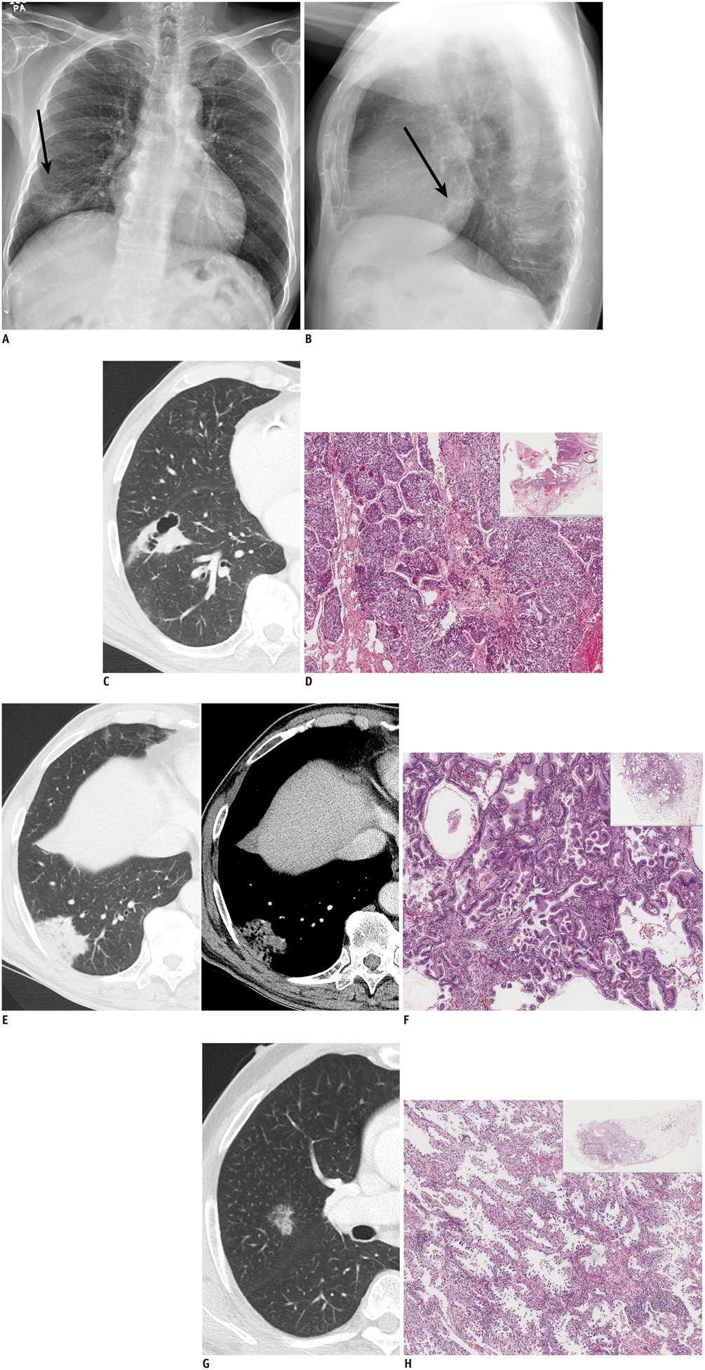
Fig. 1 72-year-old man with synchronous triple primary lung cancers. A, B. Posteroanterior (A) and lateral (B) chest radiographs reveal 4.0-cm ill-defined mass in right lower lobe (arrows). Lung window images (window level, -500 Hounsfield unit [HU]; window width, 1500 HU) of CT scan (2.5-mm slice thickness) of thorax and corresponding microscopic features at low magnification (hematoxylin and eosin stain, × 5 [× 1 in inset]). C. Anterior basal segment of right lower lobe is showing 4.2 cm irregular cavitary lesion. D. This lesion was confirmed to be squamous cell carcinoma with moderate differentiation exhibiting nests of polygonal cells with pink cytoplasm and distinct cell borders. E. 3.8-cm parenchymal low-attenuating consolidative lesion is at posterior-basal segment of right lower lobe. F. This lesion was confirmed to be invasive mucinous adenocarcinoma composed of glands lined by tall columnar mucin-containing cells. G. 2.5-cm well-defined part-solid nodular lesion is at posterior segment of right upper lobe. H. It revealed adenocarcinoma with moderate differentiation and acinar pattern showing acini of polyhedral cells.
Reference
-
1. Sulkes A, Naparstek Y, Shalit M, Kopolovic J. Second primary lung carcinoma. J Surg Oncol. 1980; 15:375–380.2. Aoki T, Tomoda Y, Watanabe H, Nakata H, Kasai T, Hashimoto H, et al. Peripheral lung adenocarcinoma: correlation of thin-section CT findings with histologic prognostic factors and survival. Radiology. 2001; 220:803–809.3. Chaudhuri MR. Primary pulmonary cavitating carcinomas. Thorax. 1973; 28:354–366.4. Lee SM, Goo JM, Park CM, Lee HJ, Im JG. A new classification of adenocarcinoma: what the radiologists need to know. Diagn Interv Radiol. 2012; 18:519–526.5. Vourtsi A, Gouliamos A, Moulopoulos L, Papacharalampous X, Chatjiioannou A, Kehagias D, et al. CT appearance of solitary and multiple cystic and cavitary lung lesions. Eur Radiol. 2001; 11:612–622.6. Ferguson MK, DeMeester TR, DesLauriers J, Little AG, Piraux M, Golomb H. Diagnosis and management of synchronous lung cancers. J Thorac Cardiovasc Surg. 1985; 89:378–385.7. Deschamps C, Pairolero PC, Trastek VF, Payne WS. Multiple primary lung cancers. Results of surgical treatment. J Thorac Cardiovasc Surg. 1990; 99:769–777. discussion 777-778.8. Wu SC, Lin ZQ, Xu CW, Koo KS, Huang OL, Xie DQ. Multiple primary lung cancers. Chest. 1987; 92:892–896.9. Ferguson MK. Synchronous primary lung cancers. Chest. 1993; 103:4 Suppl. 398S–400S.10. Huang J, Behrens C, Wistuba I, Gazdar AF, Jagirdar J. Molecular analysis of synchronous and metachronous tumors of the lung: impact on management and prognosis. Ann Diagn Pathol. 2001; 5:321–329.11. Martini N, Melamed MR. Multiple primary lung cancers. J Thorac Cardiovasc Surg. 1975; 70:606–612.12. Ostrovnaya I, Olshen AB, Seshan VE, Orlow I, Albertson DG, Begg CB. A metastasis or a second independent cancer? Evaluating the clonal origin of tumors using array copy number data. Stat Med. 2010; 29:1608–1621.13. Carey FA, Donnelly SC, Walker WS, Cameron EW, Lamb D. Synchronous primary lung cancers: prevalence in surgical material and clinical implications. Thorax. 1993; 48:344–346.14. Sarper A, Ozbilim G, Demircan A. Metachronous triple cancer: esophageal carcinoma 4 years later the synchronous bilateral bronchogenic carcinoma. Eur J Cardiothorac Surg. 2003; 24:303.15. Tokat AO, Ozkan M, Güngör A. [Synchronous lung carcinoma: a case report]. Tuberk Toraks. 2003; 51:70–73.
- Full Text Links
-
- Actions
-
Cited
- CITED
-
- Close
- Share
-
- Similar articles
-
- A case report of metachronous triple primary cancers including stomach, bladder and lung
- Synchronous Triple Primary Lung Cancer: A Rare Case with Radiologic-Pathologic Correlation
- A Case of Synchronous Triple Primary Cancer of Gastric Adenocarcinoma, Carcinoid Tumor of the Ampulla of Vater and Renal Cell Carcinoma
- Tripe synchronous primary lung cancer: one case report
- A Case of Synchronous Triple Primary Cancers Occurring in the Stomach, Colon and Liver
