Percutaneous Access via the Recanalized Paraumbilical Vein for Varix Embolization in Seven Patients
- Affiliations
-
- 1Department of Radiology, Seoul National University College of Medicine, Institute of Radiation Medicine, Seoul National University Medical Research Center, and Clinical Research Institute, Seoul National University Hospital, Seoul 110-744, Korea. angioint
- 2Department of Radiology, Dongsan Hospital, Keimyung University College of Medicine, Daegu 700-712, Korea.
- KMID: 1734948
- DOI: http://doi.org/10.3348/kjr.2014.15.5.630
Abstract
OBJECTIVE
To evaluate the feasibility of percutaneous access via the recanalized paraumbilical vein for varix embolization.
MATERIALS AND METHODS
Between July 2008 and Jan 2014, percutaneous access via the recanalized paraumbilical vein for varix embolization was attempted in seven patients with variceal bleeding. Paraumbilical vein puncture was performed under ultrasonographic guidance, followed by introduction of a 5-Fr sheath. We retrospectively evaluated the technical feasibility, procedure-related complications, and clinical outcomes of each patient.
RESULTS
Recanalized paraumbilical vein catheterization was performed successfully in all patients. Gastroesophageal varix embolization was performed in six patients, and umbilical varix embolization was performed in one patient. Embolic materials used are N-butyl cyanoacrylate (n = 6) and coil with N-butyl cyanoacrylate (n = 1). There were no procedure-related complications. One patient underwent repeated variceal embolization 6 hours after initial procedure via recanalized paraumbilical vein, due to rebleeding from gastric varix.
CONCLUSION
Percutaneous access via the paraumbilical vein for varix embolization is a simple alternative in patients with portal hypertension.
Keyword
MeSH Terms
Figure
-
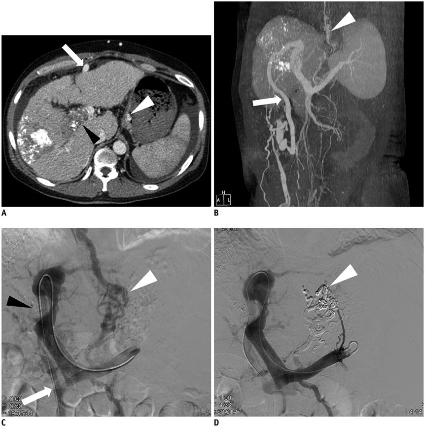
Fig. 1 52-year-old man presented with melena. A. Transverse CT scan shows tumor thrombus in right portal vein (black arrowhead), gastric varix (white arrowhead), and recanalized paraumbilical vein (arrow). B. Maximum intensity projection image of left anterior oblique view shows gastroesophageal varix (arrowhead) and recanalized paraumbilical vein (arrow). C. Splenic venography shows gastroesophageal varix (white arrowhead) and filling defect caused by tumor thrombus (black arrowhead). Note that catheter was advanced via paraumbilical vein (arrow). D. Splenic venography after variceal embolization shows subtraction artifact (arrowhead) caused by embolic materials (coils and mixture of N-butyl cyanoacrylate and iodized oil) in gastric varix.
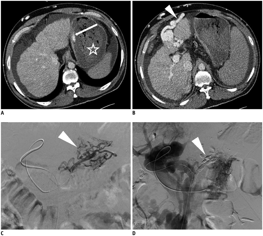
Fig. 2 49-year-old man presented with melena. A. Transverse CT scan shows extravasation of contrast media (arrow) and blood clot (star) within stomach. B. Transverse CT scan shows recanalized paraumbilical vein (arrowhead). C. Left gastric venography shows gastroesophageal varix (arrowhead). D. Splenic venography after variceal embolization shows subtraction artifact (arrowhead) caused by embolic materials (mixture of N-butyl cyanoacrylate and iodized oil).
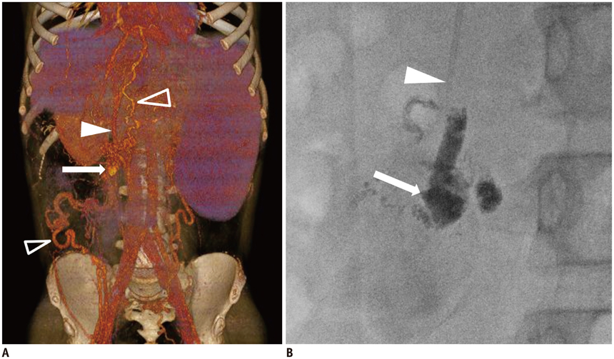
Fig. 3 11-year-old boy presented with umbilical varix bleeding. A. Volume-rendering image of CT scan shows recanalized paraumbilical vein (arrowhead), umbilical varix (arrow), and draining systemic veins (open arrowheads). B. Spot image after embolization shows embolic materials within umbilical varix (arrow) and 5-Fr sheath (arrowhead) which was inserted in paraumbilical vein.
Reference
-
1. Yamagami T, Yoshimatsu R, Miura H, Matsumoto T, Hasebe T. The role of divided injections of a sclerotic agent over two days in balloon-occluded retrograde transvenous obliteration for large gastric varices. Korean J Radiol. 2013; 14:439–445.2. Gaba RC, Omene BO, Podczerwinski ES, Knuttinen MG, Cotler SJ, Kallwitz ER, et al. TIPS for treatment of variceal hemorrhage: clinical outcomes in 128 patients at a single institution over a 12-year period. J Vasc Interv Radiol. 2012; 23:227–235.3. Kwak HS, Han YM. Percutaneous transportal sclerotherapy with N-butyl-2-cyanoacrylate for gastric varices: technique and clinical efficacy. Korean J Radiol. 2008; 9:526–533.4. Lim DH, Kim DH, Kim MS, Kim CS. Balloon-occluded percutaneous transhepatic obliteration of isolated vesical varices causing gross hematuria. Korean J Radiol. 2013; 14:94–96.5. Tian X, Wang Q, Zhang C, Liu F, Cui Y, Liu F, et al. Modified percutaneous transhepatic variceal embolization with 2-octylcyanoacrylate for bleeding gastric varices: long-term follow-up outcomes. AJR Am J Roentgenol. 2011; 197:502–509.6. Chu HH, Kim HC, Jae HJ, Yi NJ, Lee KW, Suh KS, et al. Percutaneous transsplenic access to the portal vein for management of vascular complication in patients with chronic liver disease. Cardiovasc Intervent Radiol. 2012; 35:1388–1395.7. Zhu K, Meng X, Zhou B, Qian J, Huang W, Deng M, et al. Percutaneous transsplenic portal vein catheterization: technical procedures, safety, and clinical applications. J Vasc Interv Radiol. 2013; 24:518–527.8. Lafortune M, Constantin A, Breton G, Légaré AG, Lavoie P. The recanalized umbilical vein in portal hypertension: a myth. AJR Am J Roentgenol. 1985; 144:549–553.9. Aagaard J, Jensen LI, Sørensen TI, Christensen U, Burcharth F. Recanalized umbilical vein in portal hypertension. AJR Am J Roentgenol. 1982; 139:1107–1110.10. Sparks FC, Maitem A, Glickman MG, Tilson MD. Embolization of bleeding esophageal varices via umbilical vein. An alternative approach. Arch Surg. 1982; 117:354–358.11. Spigos DG, Tauber JW, Tan WS, Mulligan BD, Espinoza GA. Work in progress: umbilical venous cannulation: a new approach for embolization of esophageal varices. Radiology. 1983; 146:53–56.12. Ibukuro K, Kojima K, Kigawa I, Tanaka R, Fukuda H, Abe S, et al. Embolization of rectal varices via a paraumbilical vein with an abdominal wall approach in a patient with massive ascites. J Vasc Interv Radiol. 2009; 20:1259–1261.13. Lim LG, Lee YM, Tan L, Chang S, Lim SG. Percutaneous paraumbilical embolization as an unconventional and successful treatment for bleeding jejunal varices. World J Gastroenterol. 2009; 15:3823–3826.14. Kim SC, Kim HC, Chung JW, Jae HJ, Park JH. Percutaneous transumbilical portal vein embolization in a patient with a ruptured hepatocellular carcinoma supplied by the portal vein. Cardiovasc Intervent Radiol. 2011; 34:Suppl 2. S214–S217.15. Lee YJ, Shin BS, Lee IH, Ohm JY, Lee BS, Ahn M, et al. Intrahepatic portosystemic venous shunt: successful embolization using the Amplatzer Vascular Plug II. Korean J Radiol. 2012; 13:827–831.16. Park KB, Choo SW, Do YS, Shin SW, Cho SG, Choo IW. Percutaneous angioplasty of portal vein stenosis that complicates liver transplantation: the mid-term therapeutic results. Korean J Radiol. 2005; 6:161–166.17. Durham JD, Kumpe DA, Van Stiegmann G, Goff JS, Subber SW, Rothbarth LJ. Direct catheterization of the mesenteric vein: combined surgical and radiologic approach to the treatment of variceal hemorrhage. Radiology. 1990; 177:229–233.18. Terabayashi H, Ohnishi K, Tsunoda T, Nakata H, Saito M, Tanaka H, et al. Prospective controlled trial of elective endoscopic sclerotherapy in comparison with percutaneous transhepatic obliteration of esophageal varices in patients with nonalcoholic cirrhosis. Gastroenterology. 1987; 93:1205–1209.
- Full Text Links
-
- Actions
-
Cited
- CITED
-
- Close
- Share
-
- Similar articles
-
- Hemoperitoneum due to Ruptured Paraumbilical Vein in a Cirrhotic Patient with Portal Hypertension: Treatment by means of Coil Embolization
- Percutaneous Embolization of the Internal Spermatic Vein for the Treatment of Childhood and Adolescent Varicocele
- A Case Report: Varix of the Intrafetal Umbilical Vein
- Percutaneous Embolization of the Internal Spermatic Vein for Treatment of Varicocele: 4 Cases
- A Case of Abdominal Wall Hematoma and Hemoperitoneum due to Rupture of Paraumbilical Vein in a Cirrhotic Patient
