Clinical Role of Interstitial Pneumonia in Patients with Scrub Typhus: A Possible Marker of Disease Severity
- Affiliations
-
- 1Department of Radiology, College of Medicine, The Catholic University of Korea, Seoul, Korea.
- 2Department of Internal Medicine, College of Medicine, The Catholic University of Korea, Seoul, Korea. cmckyo@yahoo.co.kr
- KMID: 1733506
- DOI: http://doi.org/10.3346/jkms.2004.19.5.668
Abstract
- Interstitial pneumonia (IP) frequently occurs in patients with scrub typhus, but its clinical significance is not well known. This study was designed to evaluate interstitial pneumonia as a marker of severity of the disease for patients with scrub typhus. We investigated clinical parameters representing the severity of the disease, and the chest radiographic findings for 101 patients with scrub typhus. We then compared these clinical factors between patients with and without IP. We also studied the relationship between IP and other chest radiographic findings. The chest radiography showed IP (51.4%), pleural effusion (42.6%), cardiomegaly (14.9%), pulmonary alveolar edema (20.8%), hilar lymphadenopathy (13.8%) and focal atelectasis (11.8%), respectively. The patients with IP (n=52) had higher incidences in episode of hypoxia (p=0.030), hypotension (p=0.024), severe thrombocytopenia (p=0.036) and hypoalbuminemia (p=0.013) than the patients without IP (n=49). The patients with IP also had higher incidences of pleural effusion (p<0.001), focal atelectasis (p=0.019), cardiomegaly (p<0.001), pulmonary alveolar edema (p=0.011) and hilar lymphadenopathy (p<0.001) than the patients without IP. Our data suggest that IP frequently occurs for patients with scrub typhus and its presence is closely associated with the disease severity of scrub typhus.
MeSH Terms
-
Adult
Aged
Aged, 80 and over
Female
Fluorescent Antibody Technique, Indirect
Humans
Incidence
Lung/microbiology/radiography
Lung Diseases, Interstitial/epidemiology/*microbiology/*radiography
Male
Middle Aged
Pleural Effusion/epidemiology/microbiology/radiography
Predictive Value of Tests
Prognosis
Scrub Typhus/*complications/epidemiology/*radiography
*Severity of Illness Index
Figure
-

Fig. 1 Thin-section CT through the lower lung zones in a 43-yr-old woman with scrub typhus. There is thickening of the interlobular septa and small bilateral pleural effusions.

Fig. 2 Chest radiograph and thin-section CT in a 58-yr-old woman with scrub typhus. (A) Chest radiograph shows diffuse bronchial wall thickening, diffuse ground glass opacities, mild cardiomegaly, bilateral pleural effusions and subsegmental atelectasis. (B) CT of lower zones shows interlobular septal thickening, bronchial wall thickening, diffuse ground glass opacities and patchy consolidations in the dependent lung zones, increased vascular diameter, mild cardiomegaly, bilateral pleural effusions and subsegmental atelectasis.
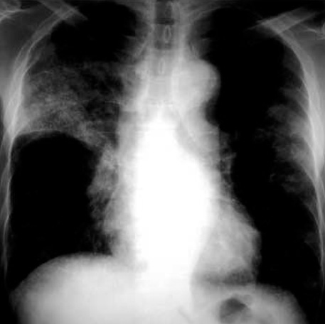
Fig. 3 Chest radiograph in a 70-yr-old man with scrub typhus. There are bilateral multiple patchy ground glass opacities and minimal right pleural effusion. The consolidations in the upper and peripheral lung zones are suggestive of ARDS.
Cited by 4 articles
-
Risk Factors for Mortality in Patients with Serratia marcescens Bacteremia
Sun Bean Kim, Yong Duk Jeon, Jung Ho Kim, Jae Kyoung Kim, Hea Won Ann, Heun Choi, Min Hyung Kim, Je Eun Song, Jin Young Ahn, Su Jin Jeong, Nam Su Ku, Sang Hoon Han, Jun Yong Choi, Young Goo Song, June Myung Kim
Yonsei Med J. 2015;56(2):348-354. doi: 10.3349/ymj.2015.56.2.348.Clinical Features of 212 Cases of Scrub Typhus in Southern Region of Gyeonggi-Do and The Significance of Initial Simple Chest X-Ray
Seong Heon Wie, U Im Chang, Hyung Wook Kim, Jian Hur, Sang IL Kim, Yang Ree Kim, Moon Won Kang
Infect Chemother. 2008;40(1):40-45. doi: 10.3947/ic.2008.40.1.40.Bell's Palsy Associated with Scrub Typhus
Sun-Young Oh, Ji-Hoon Kang, Chang-Seop Lee
Infect Chemother. 2010;42(4):253-256. doi: 10.3947/ic.2010.42.4.253.Patients with Acute Respiratory Distress Syndrome Caused by Scrub Typhus: Clinical Experiences of Eight Patients
Sun Young Kim, Hang Jea Jang, Hyunkuk Kim, Kyunghwa Shin, Mi-Hyun Kim, Kwangha Lee, Ki Uk Kim, Hye-Kyung Park, Min Ki Lee
Korean J Crit Care Med. 2014;29(3):189-193. doi: 10.4266/kjccm.2014.29.3.189.
Reference
-
1. Hwang TS, Chu YC, Kim YB, Lim BU, Kang JS. Pathologic study of mice infected with Rickettsia tsutsugamushi R19 strain. J Korean Med Sci. 1993. 8:437–445.
Article2. Watt G, Parola P. Scrub typhus and tropical rickettsioses. Curr Opin Infect Dis. 2003. 16:429–436.
Article3. Tsay RW, Chang FY. Serious complications in scrub typhus. J Microbiol Immunol Infect. 1998. 31:240–244.4. Chi WC, Huang JJ, Sung JM, Lan RR, Ko WC, Chen FF. Scrub typhus associated with multiorgan failure: a case report. Scand J Infect Dis. 1997. 29:634–635.
Article5. Kantipong P, Watt G, Jongsakul K, Choenchitra C. Infection with human immunodeficiency virus does not influence the clinical severity of scrub typhus. Clin Infect Dis. 1996. 23:1168–1170.
Article6. Seong SY, Choi MS, Kim IS. Orientia tsutsugamushi infection: overview and immune responses. Microbes Infect. 2001. 3:11–21.7. Thap LC, Supanaranond W, Treeprasertsuk S, Kitvatanachai S, Chinprasatsak S, Phonrat B. Septic shock secondary to scrub typhus: characteristics and complications. Southeast Asian J Trop Med Public Health. 2002. 33:780–786.8. Strickman D, Smith CD, Corcoran KD, Ngampochjana M, Watcharapichat P, Phulsuksombati D, Tanskul P, Dasch GA, Kelly DJ. Pathology of Rickettsia tsutsugamushi infection in Bandicota savilei, a natural host in Thailand. Am J Trop Med Hyg. 1994. 51:416–423.
Article9. Kim SJ, Chung IK, Chung TS, Song DH, Park SH, Kim HS, Lee MH. The clinical significance of upper gastrointestinal endoscopy in gastrointestinal vasculitis related to scrub typhus. Endoscopy. 2000. 32:950–955.
Article10. Sirisanthana V, Puthanakit T, Sirisanthana T. Epidemiologic, clinical and laboratory features of scrub typhus in thirty Thai children. Pediatr Infect Dis J. 2003. 22:341–345.
Article11. Choi YH, Kim SJ, Lee JY, Pai HJ, Lee KY, Lee YS. Scrub typhus; radiological and clinical findings. Clin Radiol. 2000. 55:140–144.
Article12. Kim OH, Oh DH, Kim KS, Woo JH, Kwon JH. Chest radiographic findings of scrub typhus: an analysis of 160 cases occurred in Ulsan area. J Korean Radiol Soc. 1993. 29:205–210.
Article13. Im JG, Lee KS, Kim JH, Lee WJ. Pulmonary manifestations of tsutsugamushi disease. J Korean Radiol Soc. 1988. 24:750–755.
Article14. Kim YO, Yoon SA, Ku YM, Yang CW, Kim YS, Kim SY, Choi EJ, Chang YS, Bang BK. Serum albumin level correlates with disease severity in patients with hemorrhagic fever with renal syndrome. J Korean Med Sci. 2003. 18:696–700.
Article15. Chayakul P, Panich V, Silpapojakul K. Scrub typhus pneumonitis: an entity which is frequently missed. Q J Med. 1988. 68:595–602.16. Murata K, Itoh H, Todo G, Kanaoka M, Noma S, Itoh T, Furuta M, Asamoto H, Torizuka K. Centrilobular lesions of the lung: demonstration by high resolution CT and pathologic correlation. Radiology. 1986. 161:641–645.17. Levine HD. Pathologic study of thirty-one cases of scrub typhus fever with especial reference to the cardiovascular system. Am Heart J. 1946. 31:314–328.
Article18. Yotsukura M, Aoki N, Fukuzumi N, Ishikawa K. Review of a case of tsutsugamushi disease showing myocarditis and confirmation of rickettsia by endomyocardial biopsy. Jpn Circ J. 1991. 55:149–153.
Article19. Kundin WD, Liu C, Harmon P, Rodina P. Pathogenesis of scrub typhus infection (Rickettsia tsutsugamushi) as studied by immunofluorescence. J Immunol. 1964. 93:772–781.
- Full Text Links
-
- Actions
-
Cited
- CITED
-
- Close
- Share
-
- Similar articles
-
- A Case of Scrub Typhus with Bilateral Sudden Hearing Loss
- Clinical Features and Diagnosis of Scrub Typhus
- Severe Scrub Typhus Complicated by Myofascitis Due to the Taguchi Strain of Orientia tsutsugamushi
- Antibiotic Combination Therapy for Severe Scrub Typhus: Is It Necessary?
- Pulmonary Artery Thrombosis Associated with Scrub Typhus
