Ultrastructural Studies of Gastrointestinal Stromal Tumors
- Affiliations
-
- 1Department of Pathology, Seoul National University, College of Medicine, Seoul, Korea. shparknp@plaza.snu.ac.kr
- 2Department of Pathology, Ilsan Paik Hospital, Inje University, College of Medicine, Goyang, Korea.
- 3Department of General Surgery, Ilsan Paik Hospital, Inje University, College of Medicine, Goyang, Korea.
- KMID: 1733483
- DOI: http://doi.org/10.3346/jkms.2004.19.2.234
Abstract
- Gastrointestinal stromal tumors (GISTs) are the most common mesenchymal tumors in the gastrointestinal tract (GIT). Although interstitial cells of Cajal has been suggested as origin of this tumor, the cytological and ultrastructural features of GISTs are heterogeneous and unclear. A total 10 cases of normal gastrointestinal tissue (control), 13 GISTs of the stomach (8), small intestine (3), mesocolon (1) and liver (1), and 2 gastrointestinal autonomic nervous tumor (GANT) of small intestine were ultrastructurally studied. Normal interstitial cells of Cajal (ICC) were abundantly present around the myenteric plexuses or individually scattered through the wall of GIT. ICC was characterized by slender cytoplasmic processes, well-developed endoplasmic reticulum (ER), mitochondria, Golgi apparatus, caveolae and intermediate filaments. The GISTs and GANTs had overlapping ultrastructures. The most common and important ultrastructural features of GISTs were rich villous cytoplasmic processes, dispersed intermediate filaments and abundant SER, and those of GANTs were neurosecretory granules and skenoid fibers. Compared with ICC, the GISTs and GANTs had remarkably reduced caveolae and gap junctions. Our study suggested that ultrastructural analysis gives much information to investigate lineage differentiation of neoplastic cells and make a differential diagnosis of these tumors from other mesenchymal tumors and between GISTs and GANTs.
Keyword
MeSH Terms
-
Adult
Aged
Autonomic Nervous System/*pathology/ultrastructure
Comparative Study
Cytoplasm/pathology/ultrastructure
Female
Gastrointestinal Neoplasms/*pathology/ultrastructure
Human
Immunohistochemistry
Male
Microscopy, Electron
Middle Aged
Peripheral Nervous System Neoplasms/*pathology/ultrastructure
Stromal Cells/*pathology/ultrastructure
Support, Non-U.S. Gov't
Tumor Markers, Biological
Vacuoles/pathology/ultrastructure
Figure
-
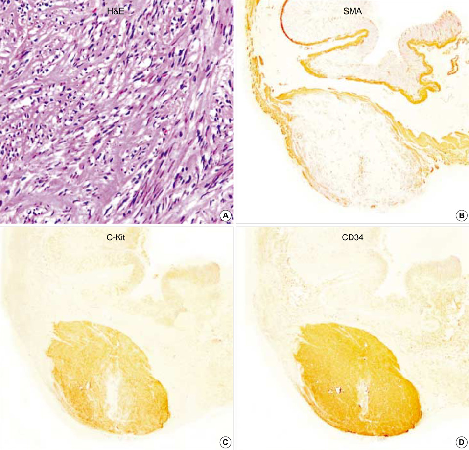
Fig. 1 Gastric GIST (case 1). The tumor is composed of interlacing fascicles of spindle cells with eosinophilic cytoplasm. The tumor cells show patch immunopositivity for SMA, and express c-Kit and CD34, strongly and diffusely. (A: H&E,×200, B: SMA, ×40, C: c-Kit, ×40, D: CD34 immunostaining, ×40).
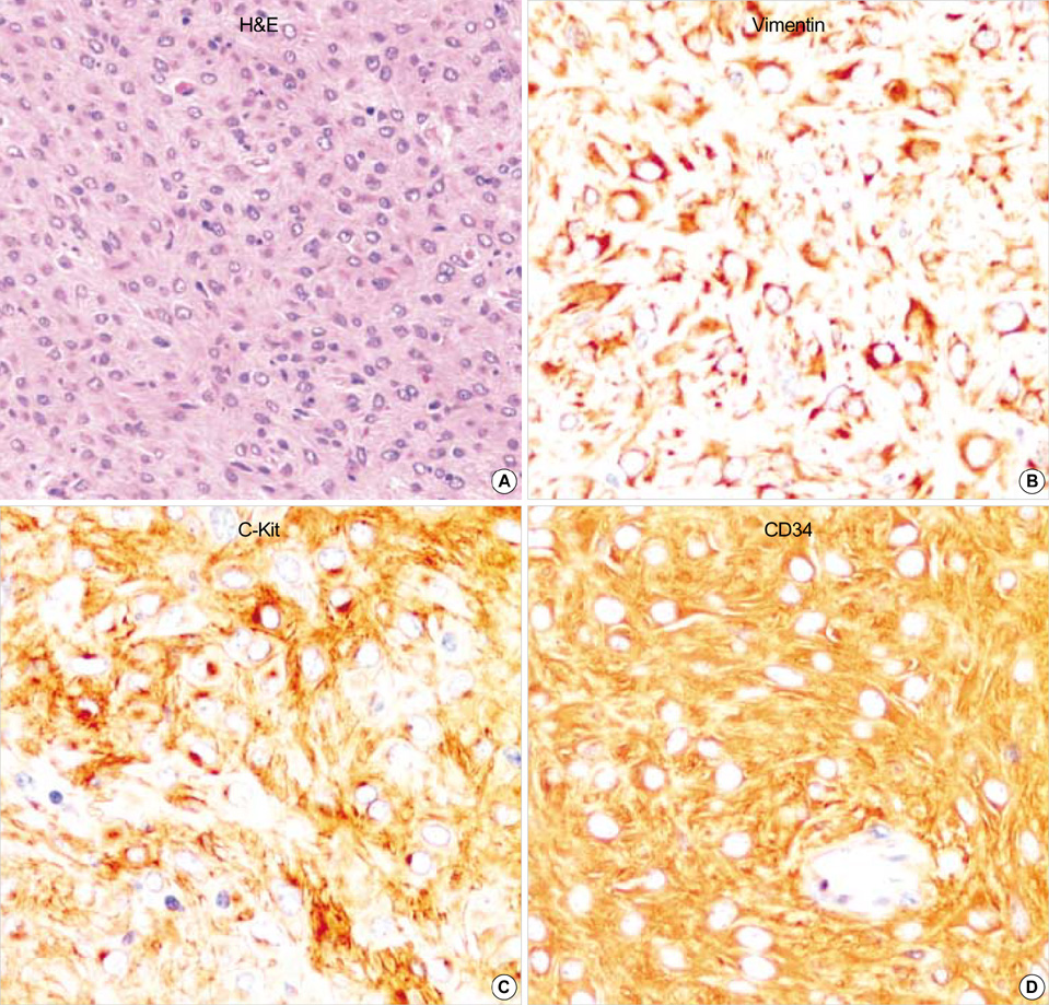
Fig. 2 Epithelioid GIST shows sheets of polygonal neoplastic cells with eosinophilic cytoplasm. This tumor reveals diffuse and strong immunoreactivity for vimentin, c-Kit and CD34. (A: H&E, ×200, B: Vimentin, ×400, C: c-Kit, ×400, D: CD34 immunostaining, ×400).
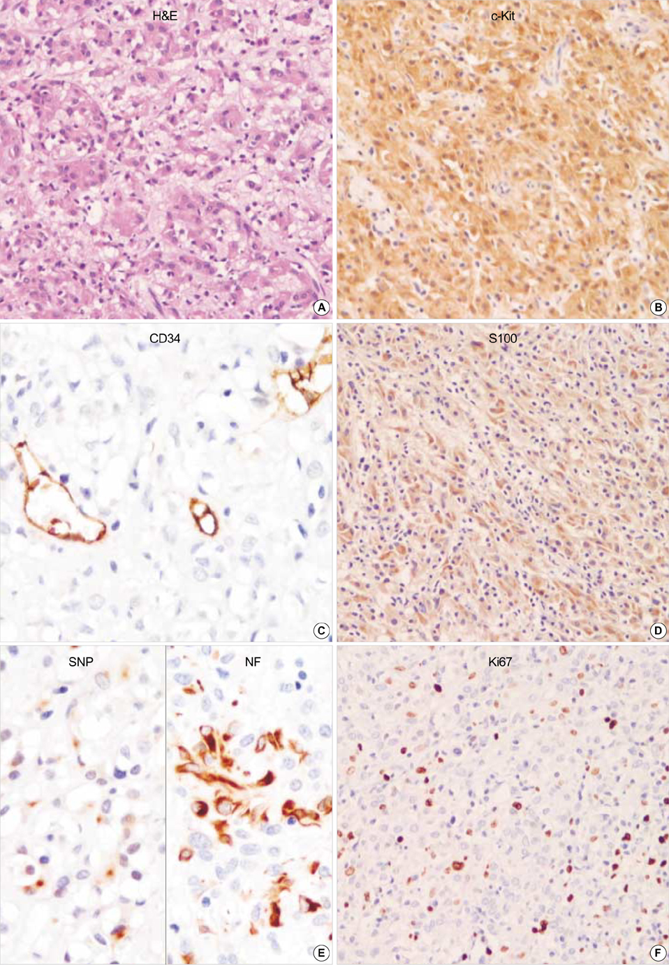
Fig. 3 GANT reveals mainly epithelioid feature. Focal cytoplasmic vacuolization is seen. The tumor cells are robustly positive for CD117 (c-kit) but negative for CD34. S-100 protein, synaptophysin and NF are partially positive in neoplastic cells. Ki67 labelling index is high. (A: H&E, ×200, B: c-Kit, ×200, C: CD34, ×400, D: S100, ×200, E: left: Synaptophysin, right: Neurofilaments, ×400, F: Ki67 immunostaining, ×200).
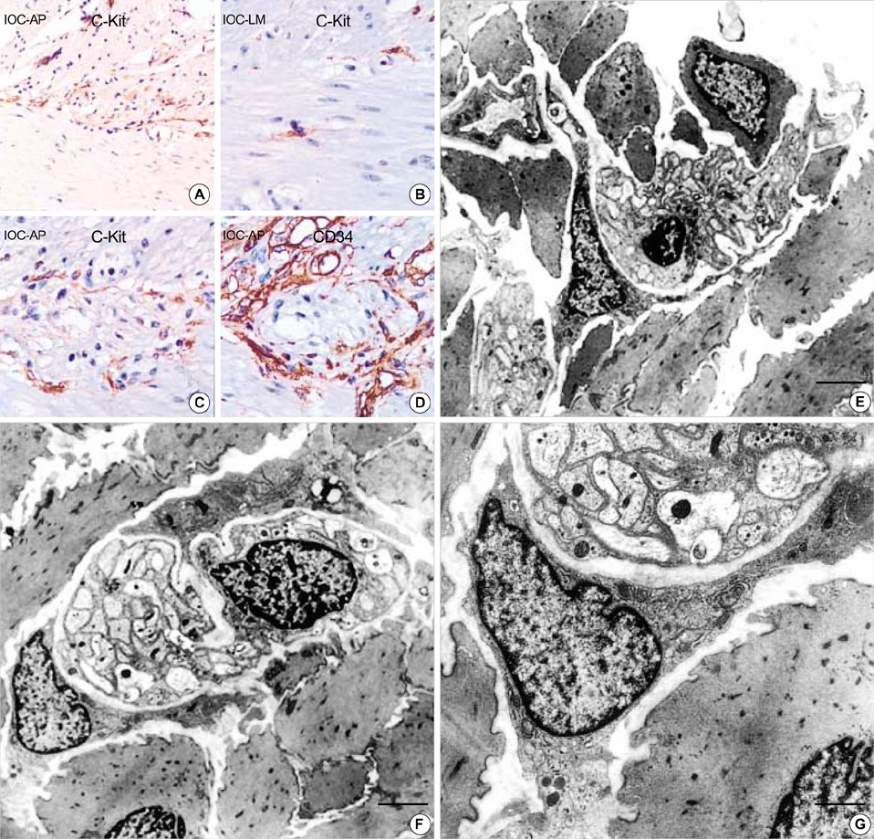
Fig. 4 (A) Normal adult (M/45 yr) small intestine. (A-D) The immunohistochemical staining for c-Kit and CD34 shows immunoreactive interstitial cell of Cajal in myenteric plexus (A: c-Kit, ×200, B: ×400, C: ×400, D: CD34, ×400). (E-G) Ultrastructure of the interstitial cell of Cajal in normal stomach. It has slender cytoplasmic processes and the nucleus is heterochromatic and lobulated in apprearance. Two interstitial cell of Cajal hug the myenteric plexuses composed of many well-delineated peripheral nerves surrounded by Schwann cell cytoplasm and continuous external lamina (F). The cytoplasm of ICC has well-defined organelles such as rough endoplasmic reticulum, mitochondria, Golgi apparatus and caveolae. Gap junctions (arrows) are present but no external lamina is noted (G). (uranyl acetate and lead citrate stain E-F: bar: 2 µm, G: bar: 1 µm, E: ×2,500, F: ×3,000, G: ×8,000).
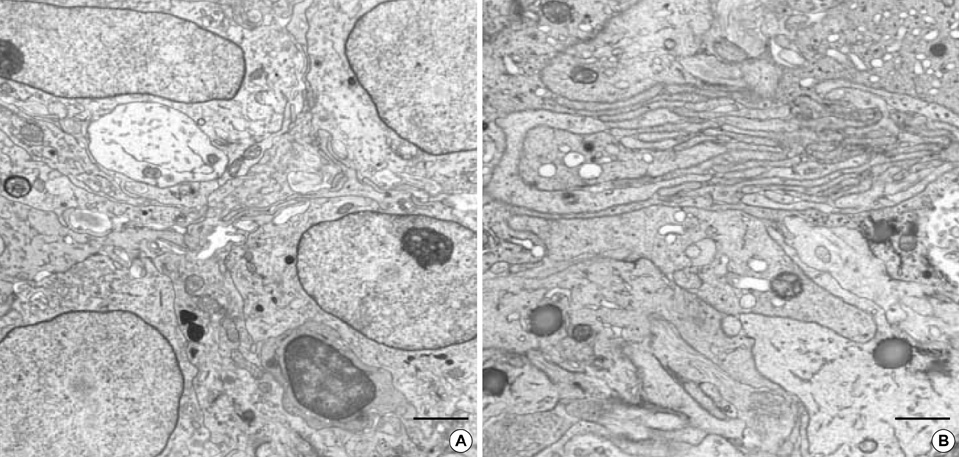
Fig. 5 The Epithelioid GIST shows euchromatic nuclei and have prominent skein appearing nucleolus with rich filopodia-like cytoplasmic processes. The cytoplasmic processes are interdigitating each other. Electron dense cytoplasmic granules, favor lysosomes, are frequently seen, which may be confused with neurosecretory granules. (uranyl acetate and lead citrate stain, A: bar: 2 µm, B: bar: 1 µm, A: ×4,000, B: ×6,000).
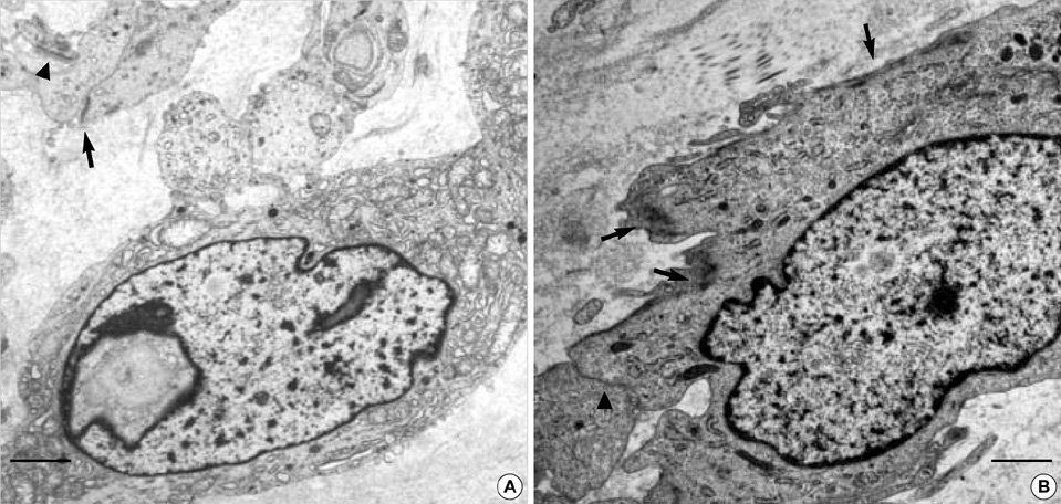
Fig. 6 (A) Spindle cell GIST also shows cytoplasmic processes but do not have much villous processes. The neoplastic cells have a nuclear pseudoinclusion and rich organelles including electron dense lysosomal granules. There are hemidesmosome-like junctions (arrowheads) as well as a few gap junctions (arrow). Incontinuous external lamina is seen. (B) Ultrastructual differentiation toward smooth muscle, such as cytoplasmic myofilaments are seen (arrows) (uranyl acetate and lead citrate stain, bar: 2 µm, A: ×5,000, B: ×6,000).
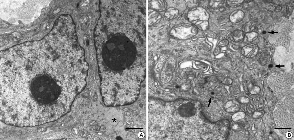
Fig. 7 The GANT have oval to irregular generally heterochromatic nuclei with prominent nucleolus. There are also abundant and evenly dispersed wavy intermediate filaments (asteric mark). The tumor cells are closely apposed except some areas having intercellular collagenous tissue. Small round neurosecretory granules (arrows) are observed, measuring 40-150 nm (uranyl acetate and lead citrate stain, A: bar: 1 µm, B: bar: 500 nm, A: ×6,000, B: ×10,000).

Fig. 8 Both cases of GANTs have characteristic extracellular skenoid fibers. (inlet: high power view of skenoid fibers). (uranyl acetate and lead citrate stain, A, B: bars: 1 µm, A, B: ×4,000).
Cited by 1 articles
-
Atopy May Be an Important Determinant of Subepithelial Fibrosis in Subjects with Asymptomatic Airway Hyperresponsiveness
Seong-Wook Sohn, Yoon-Seok Chang, Hye-Seung Lee, Doo-Hyun Chung, Choon-Taek Lee, Young-Hwan Kim, Yoon-Keun Kim, Kyung-Up Min, You-Young Kim, Sang-Heon Cho
J Korean Med Sci. 2008;23(3):390-396. doi: 10.3346/jkms.2008.23.3.390.
Reference
-
1. Chan JK. Mesenchymal tumors of the gastrointestinal tract: a paradise for acronyms (STUMP, GIST, GANT, and now GIPACT), implication of c-kit in genesis, and yet another of the many emerging roles of the interstitial cell of Cajal in the pathogenesis of gastrointestinal diseases? Adv Anat Pathol. 1999. 6:19–40.
Article2. Kindblom LG, Remotti HE, Aldenborg F, Meis-Kindblom JM. Gastrointestinal pacemaker cell tumor (GIPACT): gastrointestinal stromal tumors show phenotypic characteristics of the interstitial cells of Cajal. Am J Pathol. 1998. 152:1259–1269.3. Eyden B, Chorneyko KA, Shanks JH, Menasce LP, Banerjee SS. Contribution of electron microscopy to understanding cellular differentiation in mesenchymal tumors of the gastrointestinal tract: a study of 82 tumors. Ultrastruct Pathol. 2002. 26:269–285.
Article4. Yantiss RK, Rosenberg AE, Selig MK, Nielsen GP. Gastrointestinal stromal tumors: an ultrastructural study. Int J Surg Pathol. 2002. 10:101–113.
Article5. Liu P, Na J, Wang Y, He Q, Zhang Y, Tang X, Zou W. Study of gastrointestinal stromal tumors by light microscopy, electron microscopy and immunohistochemistry. Zhonghua Bing Li Xue Za Zhi. 2002. 31:199–203.6. Dhimes P, Lopez-Carreira M, Ortega-Serrano MP, Garcia-Munoz H, Martinez-Gonzalez MA, Ballestin C. Gastrointestinal autonomic nerve tumours and their separation from other gastrointestinal stromal tumours: an ultrastructural and immunohistochemical study of seven cases. Virchows Arch. 1995. 426:27–35.
Article7. Matsumoto K, Min W, Yamada N, Asano G. Gastrointestinal autonomic nerve tumors: immunohistochemical and ultrastructural studies in cases of gastrointestinal stromal tumor. Pathol Int. 1997. 47:308–314.
Article8. Erlandson RA, Klimstra DS, Woodruff JM. Subclassification of gastrointestinal stromal tumors based on evaluation by electron microscopy and immunohistochemistry. Ultrastruct Pathol. 1996. 20:373–393.
Article9. Montgomery E, Torbenson MS, Kaushal M, Fisher C, Abraham SC. Beta-catenin immunohistochemistry separates mesenteric fibromatosis from gastrointestinal stromal tumor and sclerosing mesenteritis. Am J Surg Pathol. 2002. 26:1296–1301.10. Makhlouf HR, Remotti HE, Ishak KG. Expression of KIT (CD117) in angiomyolipoma. Am J Surg Pathol. 2002. 26:493–497.
Article11. Leroy X, Aubert S, Leteurtre E, Gosselin B. Expression of CD117 in a malignant peripheral nerve sheath tumour arising in a patient with type 1 neurofibromatosis. Histopathology. 2003. 42:511–513.
Article12. Miettinen M, El-Rifai W, Sobin HL, Lasota J. Evaluation of malignancy and prognosis of gastrointestinal stromal tumors: a review. Hum Pathol. 2002. 33:478–483.
Article13. Mazur MT, Clark HB. Gastric stromal tumors: reappraisal of histogenesis. Am J Surg Pathol. 1983. 7:507–519.
Article14. Ma CK, Amin MB, Kintanar E, Linden MD, Zarbo RJ. Immunohistologic characterization of gastrointestinal stromal tumors: a study of 82 cases compared with 11 cases of leiomyomas. Mod Pathol. 1993. 6:139–144.15. Fletcher CD, Berman JJ, Corless C, Gorstein F, Lasota J, Longley BJ, Miettinen M, O'Leary TJ, Remotti H, Rubin BP, Shmookler B, Sobin LH, Weiss SW. Diagnosis of gastrointestinal stromal tumors: A consensus approach. Hum Pathol. 2002. 33:459–465.
Article16. Damiani S, Pasquinelli G, Eusebi V. GANT-like gastrointestinal pacemaker cell tumours with oncocytic features. Virchows Arch. 1999. 435:143–150.
Article17. Al-Bosom IA. p53 expression in gastrointestinal stromal tumors. Pathol Int. 2001. 51:519–523.
Article18. Seidal T, Edvardsson H. Expression of c-kit (CD117) and Ki67 provides information about the possible cell of origin and clinical course of gastrointestinal stromal tumours. Histopathology. 1999. 34:416–424.
Article19. Cunningham RE, Abbondanzo SL, Chu WS, Emory TS, Sobin LH, O'Leary TJ. Apoptosis, bcl-2 expression, and p53 expression in gastrointestinal stromal/smooth muscle tumors. Appl Immunohistochem Mol Morphol. 2001. 9:19–23.
Article20. Faussone-Pellegrini MS, Thuneberg L. Guide to the identification of interstitial cells of Cajal. Microsc Res Tech. 1999. 47:248–266.
Article21. Faussone-Pellegrini MS, Pantalone D, Cortesini C. An ultrastructural study of the interstitial cells of Cajal of the human stomach. J Submicrosc Cytol Pathol. 1989. 21:439–460.22. Min KW, Seo IS. Intestitial cells of Cajal in the human small intestine: immunochemical and ultrastructural study. Ultrastruct Pathol. 2003. 27:67–78.
Article23. Lee JR, Joshi V, Griffin JW Jr, Lasota J, Miettinen M. Gastrointestinal autonomic nerve tumor: immunohistochemical and molecular identity with gastrointestinal stromal tumor. Am J Surg Pathol. 2001. 25:979–987.
- Full Text Links
-
- Actions
-
Cited
- CITED
-
- Close
- Share
-
- Similar articles
-
- A case of huge Gastrointestinal stromal tumor masquerading as an ovarian malignancy
- Multiple Gastrointestinal Stromal Tumors of the Small Intestine
- A Case of Massive Bleeding from Jejunal Stromal Tumor Diagnosed by Intraoperative Enteroscopy: A Case of Jejunal Stromal Tumor Bleeding
- Clinical Relevance of the Location of Gastric Gastrointestinal Stromal Tumors
- Primary Extragastrointestinal Stromal Tumor of Retroperitoneum: Poor Response to Tyrosine Kinase Inhibitor
