Readout-Segmented Echo-Planar Imaging in Diffusion-Weighted MR Imaging in Breast Cancer: Comparison with Single-Shot Echo-Planar Imaging in Image Quality
- Affiliations
-
- 1Department of Radiology, Seoul St. Mary's Hospital, College of Medicine, The Catholic University of Korea, Seoul 137-701, Korea. rad-ksh@catholic.ac.kr
- 2Department of Radiology, Incheon St. Mary's Hospital, College of Medicine, The Catholic University of Korea, Incheon 403-720, Korea.
- 3Department of Radiology, St. Paul Hospital, College of Medicine, The Catholic University of Korea, Seoul 130-709, Korea.
- 4Healthcare Sector, Siemens Ltd., Seoul 120-837, Korea.
- 5Healthcare Sector, Siemens AG, Erlangen 91050, Germany.
- 6Department of General Surgery, Seoul St. Mary's Hospital, College of Medicine, The Catholic University of Korea, Seoul 137-701, Korea.
- KMID: 1731042
- DOI: http://doi.org/10.3348/kjr.2014.15.4.403
Abstract
OBJECTIVE
The purpose of this study was to compare the image quality of standard single-shot echo-planar imaging (ss-EPI) and that of readout-segmented EPI (rs-EPI) in patients with breast cancer.
MATERIALS AND METHODS
Seventy-one patients with 74 breast cancers underwent both ss-EPI and rs-EPI. For qualitative comparison of image quality, three readers independently assessed the two sets of diffusion-weighted (DW) images. To evaluate geometric distortion, a comparison was made between lesion lengths derived from contrast enhanced MR (CE-MR) images and those obtained from the corresponding DW images. For assessment of image parameters, signal-to-noise ratio (SNR), lesion contrast, and contrast-to-noise ratio (CNR) were calculated.
RESULTS
The rs-EPI was superior to ss-EPI in most criteria regarding the qualitative image quality. Anatomical structure distinction, delineation of the lesion, ghosting artifact, and overall image quality were significantly better in rs-EPI. Regarding the geometric distortion, lesion length on ss-EPI was significantly different from that of CE-MR, whereas there were no significant differences between CE-MR and rs-EPI. The rs-EPI was superior to ss-EPI in SNR and CNR.
CONCLUSION
Readout-segmented EPI is superior to ss-EPI in the aspect of image quality in DW MR imaging of the breast.
MeSH Terms
-
Adult
Aged
Aged, 80 and over
Artifacts
Breast/pathology
Breast Neoplasms/pathology
Contrast Media/diagnostic use
Diffusion Magnetic Resonance Imaging/methods/*standards
Echo-Planar Imaging/methods/*standards
Female
Humans
Image Enhancement/methods/*standards
Middle Aged
Observer Variation
Retrospective Studies
Sensitivity and Specificity
Signal-To-Noise Ratio
Contrast Media
Figure
-

Fig. 1 MR images in 39-year-old woman with invasive ductal carcinoma in her right breast. Contrast-enhanced T1-weighted MR image (A), readout-segmented echo-planar image (B), and ADC map (C). Lesion ROI was drawn in center of mass with high signal intensity in DW image then it was copied to ADC map. Normal tissue ROI was drawn in homogeneous breast parenchyma in contralateral breast. Another ROI was drawn at periphery of DW image to measure background noise. SNR, contrast, CNR, and lesion ADC were 304.5, 2.6, 3.4, and 1.06 × 10⁻³ mm2/sec, respectively. ADC = apparent diffusion coefficient, CNR = contrast-to-noise ratio, DW = diffusion-weighted, ROI = regions-of-interest, SNR = signal-to-noise ratio

Fig. 2 MR images in 45-year-old woman with two malignant masses in her right breast (invasive ductal carcinoma). Contrast-enhanced T1-weighted MR image (A), single-shot echo-planar image (ss-EPI) (B), and readout-segmented echo-planar image (rs-EPI) (C). While posteriorly located rim-enhancing mass is relatively well identifiable on both sequences, other larger mass is poorly defined on ss-EPI. Average scores given by three readers for overall image quality were 2.3 (poor) for ss-EPI and 5.3 (above average) for rs-EPI. Other variables are scored as follows (ss-EPI vs. rs-EPI); anatomical structure, 2.0 vs. 4.0; delineation of lesion, 2.7 vs. 5.7; ghosting artifacts, 2.7 vs. 3.0; fat suppression, 3.0 vs. 3.0; SNR, 78.0 vs. 232.0; contrast, 1.0 vs. 2.2; CNR, 0.1 vs. 3.8; ADC, 1.35 × 10⁻³ mm2/sec vs. 1.33 × 10⁻³ mm2/sec. ADC = apparent diffusion coefficient, CNR = contrast-to-noise ratio, SNR = signal-to-noise ratio

Fig. 3 Comparison of average lesion lengths in contrast-enhanced (CE)-MR and diffusion-weighted (DW) images. Both average anterior-posterior (AP) length (A) and left-right (LR) width (B) of lesions were measured smaller in both sets of DW images. Whereas there were no significant differences in AP length and LR width between CE-MR and readout-segmented echo-planar image (rs-EPI), they were significantly smaller in single-shot echo-planar image (ss-EPI).
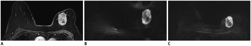
Fig. 4 MR images in 88-year-old woman with invasive ductal carcinoma in her left breast. Contrast-enhanced T1-weighted MR image (A), single-shot echo-planar image (ss-EPI) (B), and readout-segmented echo-planar image (rs-EPI) (C). rs-EPI provides superior anatomical detail and lesion delineation. Spatial distortion is more prominent on ss-EPI. Average scores given by three readers for overall image quality were 3 (acceptable) for ss-EPI and 5 (above average) for rs-EPI. Other variables are scored as follows (ss-EPI vs. rs-EPI); anatomical structure, 0 vs. 4.0; delineation of lesion, 4.3 vs. 6.0; ghosting artifacts, 2.7 vs. 4.0; fat suppression, 1.3 vs. 2.0; SNR, 126.5 vs. 556.9; contrast, 3.6 vs. 8.8; CNR, 6.3 vs. 15.4; ADC, 0.62 × 10⁻³ mm2/sec vs. 0.63 × 10⁻³ mm2/sec. ADC = apparent diffusion coefficient, CNR = contrast-to-noise ratio, SNR = signal-to-noise ratio

Fig. 5 Correlation for lesion length between contrast-enhanced (CE) T1-weighted MRI and diffusion-weighted images (DWI). Pearson correlation coefficients of anterior-posterior (AP) length (A) and left-right (LR) width (B) are slightly higher in readout-segmented echo-planar image (r = 0.995, r = 0.994) than in single-shot echo-planar image (r = 0.973, r = 0.975). rs-EPI = readout-segmented echo-planar imaging, ss-EPI = single-shot echo-planar imaging
Cited by 1 articles
-
Thyroid-Associated Orbitopathy: Evaluating Microstructural Changes of Extraocular Muscles and Optic Nerves Using Readout-Segmented Echo-Planar Imaging-Based Diffusion Tensor Imaging
Huan-Huan Chen, Hao Hu, Wen Chen, Dai Cui, Xiao-Quan Xu, Fei-Yun Wu, Tao Yang
Korean J Radiol. 2020;21(3):332-340. doi: 10.3348/kjr.2019.0053.
Reference
-
1. Huang W, Fisher PR, Dulaimy K, Tudorica LA, O'Hea B, Button TM. Detection of breast malignancy: diagnostic MR protocol for improved specificity. Radiology. 2004; 232:585–591.2. Kuhl C. The current status of breast MR imaging. Part I. Choice of technique, image interpretation, diagnostic accuracy, and transfer to clinical practice. Radiology. 2007; 244:356–378.3. Kuhl CK. Current status of breast MR imaging. Part 2. Clinical applications. Radiology. 2007; 244:672–691.4. Bluemke DA, Gatsonis CA, Chen MH, DeAngelis GA, DeBruhl N, Harms S, et al. Magnetic resonance imaging of the breast prior to biopsy. JAMA. 2004; 292:2735–2742.5. Bogner W, Gruber S, Pinker K, Grabner G, Stadlbauer A, Weber M, et al. Diffusion-weighted MR for differentiation of breast lesions at 3.0 T: how does selection of diffusion protocols affect diagnosis? Radiology. 2009; 253:341–335.6. Ei Khouli RH, Jacobs MA, Mezban SD, Huang P, Kamel IR, Macura KJ, et al. Diffusion-weighted imaging improves the diagnostic accuracy of conventional 3.0-T breast MR imaging. Radiology. 2010; 256:64–67.7. Guo Y, Cai YQ, Cai ZL, Gao YG, An NY, Ma L, et al. Differentiation of clinically benign and malignant breast lesions using diffusion-weighted imaging. J Magn Reson Imaging. 2002; 16:172–178.8. Yeom KW, Holdsworth SJ, Van AT, Iv M, Skare S, Lober RM, et al. Comparison of readout-segmented echo-planar imaging (EPI) and single-shot EPI in clinical application of diffusion-weighted imaging of the pediatric brain. AJR Am J Roentgenol. 2013; 200:W437–W443.9. Farzaneh F, Riederer SJ, Pelc NJ. Analysis of T2 limitations and off-resonance effects on spatial resolution and artifacts in echo-planar imaging. Magn Reson Med. 1990; 14:123–139.10. Porter DA, Heidemann RM. High resolution diffusion-weighted imaging using readout-segmented echo-planar imaging, parallel imaging and a two-dimensional navigator-based reacquisition. Magn Reson Med. 2009; 62:468–475.11. Robson MD, Anderson AW, Gore JC. Diffusion-weighted multiple shot echo planar imaging of humans without navigation. Magn Reson Med. 1997; 38:82–88.12. Porter DA, Mueller E. Multi-shot diffusion-weighted EPI with readout mosaic segmentation and 2D navigator correction. Proc Intl Soc Mag Reson Med. 2004; 11:442.13. Miller KL, Pauly JM. Nonlinear phase correction for navigated diffusion imaging. Magn Reson Med. 2003; 50:343–353.14. Bogner W, Pinker-Domenig K, Bickel H, Chmelik M, Weber M, Helbich TH, et al. Readout-segmented echo-planar imaging improves the diagnostic performance of diffusion-weighted MR breast examinations at 3.0 T. Radiology. 2012; 263:64–67.15. Holdsworth SJ, Yeom K, Skare S, Gentles AJ, Barnes PD, Bammer R. Clinical application of readout-segmented- echo-planar imaging for diffusion-weighted imaging in pediatric brain. AJNR Am J Neuroradiol. 2011; 32:1274–1279.16. Morelli J, Porter D, Ai F, Gerdes C, Saettele M, Feiweier T, et al. Clinical evaluation of single-shot and readout-segmented diffusion-weighted imaging in stroke patients at 3 T. Acta Radiol. 2013; 54:299–306.
- Full Text Links
-
- Actions
-
Cited
- CITED
-
- Close
- Share
-
- Similar articles
-
- Analysis of Apparent Diffusion Coefficients of the Brain in Healthy Controls: A Comparison Study between Single-Shot Echo-Planar Imaging and Read-out-Segmented Echo-Planar Imaging
- Multi-slice Multi-echo Pulsed-gradient Spin-echo (MePGSE) Sequence for Diffusion Tensor Imaging MRI: A Preliminary Result
- Ultrafast Magnetic Resonance Imaging: Echo Planar Imaging and Spiral Scan Imaging
- Single-Shot Echo-Planar Diffusion-Weighted MR Imaging at 3T and 1.5T for Differentiation of Benign Vertebral Fracture Edema and Tumor Infiltration
- Comparison of FSE and EPI with Brain MR Imaging
