The normal electroretinogram in adult healthy Shih Tzu dogs using the HMsERG
- Affiliations
-
- 1Department of Veterinary Surgery, College of Veterinary Medicine, Konkuk University, Seoul 143-701, Korea. swjeong@konkuk.ac.kr
- 2Department of Veterinary Medicine, Madingley Road, Cambridge CB3 0ES, United Kingdom.
- KMID: 1726923
- DOI: http://doi.org/10.4142/jvs.2009.10.3.233
Abstract
- Electroretinography (ERG) is a reliable diagnostic tool for the diagnosis of retinal disease. It measures electric potentials occurring in the retina in response to light stimulation. In this study, we examined the normal electroretinogram using the Handheld Multispecies ERG (HMsERG) in Shih Tzu dogs. ERG recordings were performed in twelve eyes of six healthy Shih Tzu dogs. Dogs were anesthetized with a combination of medetomidine and ketamine. Proparacaine eye drops were also applied as a topical anesthetic. Tropicamide eye drops were applied for mydriasis. After 20 min of dark adaptation, we recorded the amplitudes and implicit times of the b-waves of the rod, standard rod and cone (Std R&C), high-intensity rod and cone (Hi-int R&C), and cone systems, and responses of the cones and inner retina by flicker light stimulation (cone flicker). Results showed that mean the amplitudes of a-waves of Std R&C, Hi-int R&C, and the cone responses were 141.25 microV, 173.00 microV, and 12.92 microV, respectively. The b-waves of the rod responses ranged from 141.58 to 155.25 microV; the Std R&C was 314.75 microV, the Hi-int R&C was 329.42 microV, the cones were 37.75 microV, and the flicker responses were 64.08 microV. The b/a ratios for the Std R&C, Hi-int R&C, and the cone response were 2.29, 1.94, and 3.71, respectively. Mean implicit time of the a-wave of the Std R&C was 15.12 ms, of Hi-int R&C was 13.42 ms, and of the cone response was 7.22 ms. The b-wave of the rod responses ranged from 68.12 to 72.68 ms, of Std R&C were 37.28 ms, of Hi-int R&C were 41.90, of the cone responses were 38.12 ms, and of the cone flicker responses were 22.80 ms. We believe that these parameters can be used as reference "normal" ERGs ranges for Shih Tzu dogs using the HMsERG under medetomidine and ketamine anesthesia.
Keyword
MeSH Terms
Figure
-
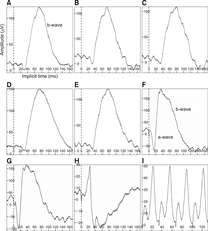
Fig. 1 Representative waveforms of the ERG results using the dog diagnostic protocol of the Handheld Multispecies electroretinography (HMsERG) in adult Shih Tzu dogs showing normal retinal function. (A-E: rod response, F: standard rod and cone (Std R&C) responses, G: high-intensity (Hi-int) R&C responses, H: cone response, I: cone flicker response).
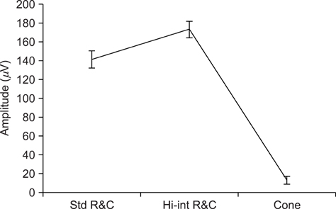
Fig. 2 a-wave amplitudes (mean ± SE) using the diagnostic protocol with the HMsERG equipment in adult Shih Tzu dogs showing normal vision. Std R&C: standard rod and cone, Hi-int R&C: high-intensity rod and cone.

Fig. 3 b-wave amplitudes (mean ± SE) using the dog diagnostic protocol with the HMsERG equipment in adult Shih Tzu dogs showing normal vision. Std R&C: standard rod and cone, Hi-int R&C: high-intensity rod and cone.

Fig. 4 b/a ratio of amplitudes (mean ± SE) using dog diagnostic protocol of HMsERG equipment in adult Shih Tzu dogs showing normal vision. Std R&C: standard rod and cone, Hi-int R&C: high-intensity rod and cone.
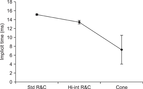
Fig. 5 a-wave implicit time (mean ± SE) using dog diagnostic protocol of HMsERG equipment in adult Shih Tzu dogs showing normal vision. Std R&C: standard rod and cone, Hi-int R&C: high-intensity rod and cone.

Fig. 6 b-wave implicit time (mean ± SE) using dog diagnostic protocol of HMsERG equipment in adult Shih Tzu dogs showing normal vision. Std R&C: standard rod and cone, Hi-int R&C: high-intensity rod and cone.
Reference
-
1. Ford M, Bragadóttir R, Rakoczy PE, Narfström K. Gene transfer in the RPE65 null mutation dog: relationship between construct volume, visual behavior and electroretinographic (ERG) results. Doc Ophthalmol. 2003. 107:79–86.2. Gum GG. Electrophysiology in veterinary ophthalmology. Vet Clin North Am Small Anim Pract. 1980. 10:437–454.
Article3. Kommonen B, Raitta C. Electroretinography in Labrador Retrievers given ketamine-xylazine anesthesia. Am J Vet Res. 1987. 48:1325–1331.4. Krill AE. The electroretinogram and electro-oculogram: clinical applications. Invest Ophthalmol. 1970. 9:600–617.5. Maehara S, Itoh N, Itoh Y, Wakaiki S, Tsuzuki K, Seno T, Kushiro T, Yamashita K, Izumisawa Y, Kotani T. Electroretinography using Contact Lens Electrode with Built-in Light Source in dogs. J Vet Med Sci. 2005. 67:509–514.
Article6. Marmor MF, Holder GE, Seeliger MW, Yamamoto S. Standard for clinical electroretinography (2004 update). Doc Ophthalmol. 2004. 108:107–114.
Article7. Marmor MF, Zrenner E. Standard for clinical electroretinography (1999 update). International Society for Clinical Electrophysiology of Vision. Doc Ophthalmol. 1999. 97:143–156.8. Mentzer AE, Eifler DM, Montiani-Ferreira F, Tuntivanich N, Forcier JQ, Petersen-Jones SM. Influence of recording electrode type and reference electrode position on the canine electroretinogram. Doc Ophthalmol. 2005. 111:95–106.
Article9. Narfström K, Ekesten B, Rosolen SG, Spiess BM, Percicot CL, Ofri R. Guidelines for clinical electroretinography in the dog. Doc Ophthalmol. 2002. 105:83–92.10. Sims MH, Brooks DE. Changes in oscillatory potentials in the canine electroretinogram during dark adaptation. Am J Vet Res. 1990. 51:1580–1586.11. Slatter DH. Fundamentals of Veterinary Ophthalmology. 2001. 3rd ed. Philadelphia: Saunders;419–456.12. Tuntivanich N, Mentzer AL, Eifler DM, Montiani-Ferreira F, Forcier JQ, Johnson CA, Petersen-Jones SM. Assessment of the dark-adaptation time required for recovery of electroretinographic responses in dogs after fundus photography and indirect ophthalmoscopy. Am J Vet Res. 2005. 66:1798–1804.
Article13. Whitley RD, Vygantas KR. Martin CL, editor. Presumed inherited ocular diseases. Ophthalmic Disease in Veterinary Medicine. 2005. 1st ed. London: Manson;471–491.14. Yanase J, Ogawa H. Effects of halothane and sevoflurane on the electroretinogram of dogs. Am J Vet Res. 1997. 58:904–909.15. Yanase J, Ogawa H, Ohtsuka H. Scotopic threshold response of the electroretinogram of dogs. Am J Vet Res. 1996. 57:361–366.
- Full Text Links
-
- Actions
-
Cited
- CITED
-
- Close
- Share
-
- Similar articles
-
- Comparison of radiographic and computed tomographic acetabular index in small-breed dogs: a preliminary study using Maltese and Shih Tzu
- Delayed periocular dermatitis as a rare side-effect of topical antiglaucoma eyedrop instillation in two Shih-Tzu dogs with atopic dermatitis
- Mammary gland tumors in three male dogs
- Candida albicans urinary tract infection in a Shih Tzu dog with immune-mediated hemolytic anemia
- Application of mycophenolate mofetil for immune-mediated hemolytic anemia in two dogs
