Korean J Radiol.
2013 Aug;14(4):683-691. 10.3348/kjr.2013.14.4.683.
A Comparison of Two Commercial Volumetry Software Programs in the Analysis of Pulmonary Ground-Glass Nodules: Segmentation Capability and Measurement Accuracy
- Affiliations
-
- 1Department of Radiology, Seoul National University College of Medicine, and Institute of Radiation Medicine, Seoul National University Medical Research Center, Seoul 110-744, Korea. cmpark@radiol.snu.ac.kr
- 2Cancer Research Institute, Seoul National University, Seoul 110-744, Korea.
- KMID: 1715775
- DOI: http://doi.org/10.3348/kjr.2013.14.4.683
Abstract
OBJECTIVE
To compare the segmentation capability of the 2 currently available commercial volumetry software programs with specific segmentation algorithms for pulmonary ground-glass nodules (GGNs) and to assess their measurement accuracy.
MATERIALS AND METHODS
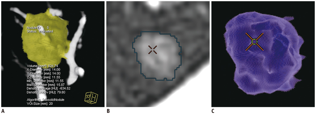
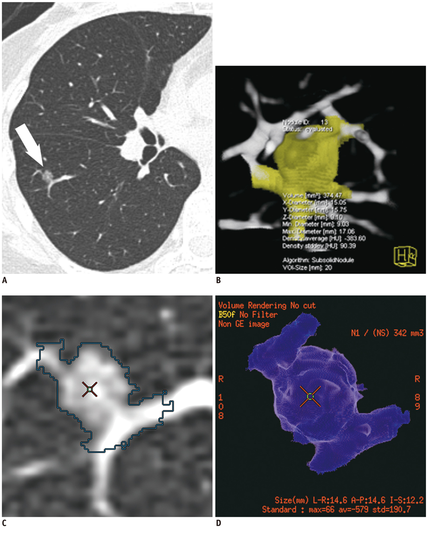
In this study, 55 patients with 66 GGNs underwent unenhanced low-dose CT. GGN segmentation was performed by using 2 volumetry software programs (LungCARE, Siemens Healthcare; LungVCAR, GE Healthcare). Successful nodule segmentation was assessed visually and morphologic features of GGNs were evaluated to determine factors affecting segmentation by both types of software. In addition, the measurement accuracy of the software programs was investigated by using an anthropomorphic chest phantom containing simulated GGNs.
RESULTS
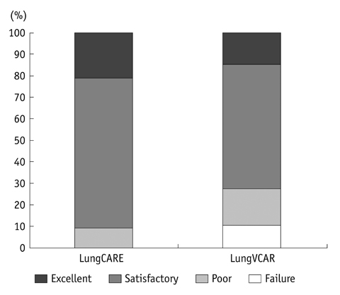
The successful nodule segmentation rate was significantly higher in LungCARE (90.9%) than in LungVCAR (72.7%) (p = 0.012). Vascular attachment was a negatively influencing morphologic feature of nodule segmentation for both software programs. As for measurement accuracy, mean relative volume measurement errors in nodules > or = 10 mm were 14.89% with LungCARE and 19.96% with LungVCAR. The mean relative attenuation measurement errors in nodules > or = 10 mm were 3.03% with LungCARE and 5.12% with LungVCAR.
CONCLUSION
LungCARE shows significantly higher segmentation success rates than LungVCAR. Measurement accuracy of volume and attenuation of GGNs is acceptable in GGNs > or = 10 mm by both software programs.
Keyword
MeSH Terms
Figure
Cited by 2 articles
-
Quality of Radiomic Features in Glioblastoma Multiforme: Impact of Semi-Automated Tumor Segmentation Software
Myungeun Lee, Boyeong Woo, Michael D. Kuo, Neema Jamshidi, Jong Hyo Kim
Korean J Radiol. 2017;18(3):498-509. doi: 10.3348/kjr.2017.18.3.498.Imaging Informatics: A New Horizon for Radiologyin the Era of Artificial Intelligence, Big Data, and Data Science
Jong Hyo Kim
J Korean Soc Radiol. 2019;80(2):176-201. doi: 10.3348/jksr.2019.80.2.176.
Reference
-
1. Henschke CI, Yankelevitz DF, Mirtcheva R, McGuinness G, McCauley D, Miettinen OS. ELCAP Group. CT screening for lung cancer: frequency and significance of part-solid and nonsolid nodules. AJR Am J Roentgenol. 2002; 178:1053–1057.2. Park CM, Goo JM, Lee HJ, Lee CH, Chun EJ, Im JG. Nodular ground-glass opacity at thin-section CT: histologic correlation and evaluation of change at follow-up. Radiographics. 2007; 27:391–408.3. Lee HJ, Goo JM, Lee CH, Park CM, Kim KG, Park EA, et al. Predictive CT findings of malignancy in ground-glass nodules on thin-section chest CT: the effects on radiologist performance. Eur Radiol. 2009; 19:552–560.4. Kim HY, Shim YM, Lee KS, Han J, Yi CA, Kim YK. Persistent pulmonary nodular ground-glass opacity at thin-section CT: histopathologic comparisons. Radiology. 2007; 245:267–275.5. Tsunezuka Y, Shimizu Y, Tanaka N, Takayanagi T, Kawano M. Positron emission tomography in relation to Noguchi's classification for diagnosis of peripheral non-small-cell lung cancer 2 cm or less in size. World J Surg. 2007; 31:314–317.6. de Hoop B, Gietema H, van de Vorst S, Murphy K, van Klaveren RJ, Prokop M. Pulmonary ground-glass nodules: increase in mass as an early indicator of growth. Radiology. 2010; 255:199–206.7. Goo JM. A computer-aided diagnosis for evaluating lung nodules on chest CT: the current status and perspective. Korean J Radiol. 2011; 12:145–155.8. Oda S, Awai K, Murao K, Ozawa A, Yanaga Y, Kawanaka K, et al. Computer-aided volumetry of pulmonary nodules exhibiting ground-glass opacity at MDCT. AJR Am J Roentgenol. 2010; 194:398–406.9. Park CM, Goo JM, Lee HJ, Kim KG, Kang MJ, Shin YH. Persistent pure ground-glass nodules in the lung: interscan variability of semiautomated volume and attenuation measurements. AJR Am J Roentgenol. 2010; 195:W408–W414.10. de Hoop B, Gietema H, van Ginneken B, Zanen P, Groenewegen G, Prokop M. A comparison of six software packages for evaluation of solid lung nodules using semi-automated volumetry: what is the minimum increase in size to detect growth in repeated CT examinations. Eur Radiol. 2009; 19:800–808.11. Kakinuma R, Kodama K, Yamada K, Yokoyama A, Adachi S, Mori K, et al. Performance evaluation of 4 measuring methods of ground-glass opacities for predicting the 5-year relapse-free survival of patients with peripheral nonsmall cell lung cancer: a multicenter study. J Comput Assist Tomogr. 2008; 32:792–798.12. Wang Y, van Klaveren RJ, van der Zaag-Loonen HJ, de Bock GH, Gietema HA, Xu DM, et al. Effect of nodule characteristics on variability of semiautomated volume measurements in pulmonary nodules detected in a lung cancer screening program. Radiology. 2008; 248:625–631.13. Gavrielides MA, Kinnard LM, Myers KJ, Petrick N. Noncalcified lung nodules: volumetric assessment with thoracic CT. Radiology. 2009; 251:26–37.14. Kostis WJ, Reeves AP, Yankelevitz DF, Henschke CI. Three-dimensional segmentation and growth-rate estimation of small pulmonary nodules in helical CT images. IEEE Trans Med Imaging. 2003; 22:1259–1274.15. Das M, Ley-Zaporozhan J, Gietema HA, Czech A, Mühlenbruch G, Mahnken AH, et al. Accuracy of automated volumetry of pulmonary nodules across different multislice CT scanners. Eur Radiol. 2007; 17:1979–1984.16. Petrou M, Quint LE, Nan B, Baker LH. Pulmonary nodule volumetric measurement variability as a function of CT slice thickness and nodule morphology. AJR Am J Roentgenol. 2007; 188:306–312.17. Godoy MC, Naidich DP. Subsolid pulmonary nodules and the spectrum of peripheral adenocarcinomas of the lung: recommended interim guidelines for assessment and management. Radiology. 2009; 253:606–622.18. Travis WD, Brambilla E, Noguchi M, Nicholson AG, Geisinger KR, Yatabe Y, et al. International association for the study of lung cancer/american thoracic society/european respiratory society international multidisciplinary classification of lung adenocarcinoma. J Thorac Oncol. 2011; 6:244–285.19. Warfield SK, Zou KH, Wells WM. Simultaneous truth and performance level estimation (STAPLE): an algorithm for the validation of image segmentation. IEEE Trans Med Imaging. 2004; 23:903–921.20. Deeley MA, Chen A, Datteri R, Noble JH, Cmelak AJ, Donnelly EF, et al. Comparison of manual and automatic segmentation methods for brain structures in the presence of space-occupying lesions: a multi-expert study. Phys Med Biol. 2011; 56:4557–4577.
- Full Text Links
- Actions
-
Cited
- CITED
-
- Close
- Share
- Similar articles
-
- Effect of the High-Pitch Mode in Dual-Source Computed Tomography on the Accuracy of Three-Dimensional Volumetry of Solid Pulmonary Nodules: A Phantom Study
- A New Method of Measuring the Amount of Soft Tissue in Pulmonary Ground-Glass Opacity Nodules: a Phantom Study
- Volumetry of Artificial Pulmonary Nodules in Ex Vivo Porcine Lungs: Comparison of Semi-automated Volumetry and Radiologists' Performance
- Evaluation of Computer Aided Volumetry for Simulated Small Pulmonary Nodules on Computed Tomography
- Pulmonary Subsolid Nodules: An Overview & Management Guidelines


