Effect of the High-Pitch Mode in Dual-Source Computed Tomography on the Accuracy of Three-Dimensional Volumetry of Solid Pulmonary Nodules: A Phantom Study
- Affiliations
-
- 1Department of Radiology, Korea University Anam Hospital, Seoul 136-705, Korea. yuwhan@kumc.or.kr
- 2Department of Radiology, Korea University Guro Hospital, Seoul 152-703, Korea.
- 3Department of Radiology, Korea University Ansan Hospital, Ansan 425-707, Korea.
- KMID: 2155535
- DOI: http://doi.org/10.3348/kjr.2015.16.3.641
Abstract
OBJECTIVE
To evaluate the influence of high-pitch mode (HPM) in dual-source computed tomography (DSCT) on the accuracy of three-dimensional (3D) volumetry for solid pulmonary nodules.
MATERIALS AND METHODS
A lung phantom implanted with 45 solid pulmonary nodules (n = 15 for each of 4-mm, 6-mm, and 8-mm in diameter) was scanned twice, first in conventional pitch mode (CPM) and then in HPM using DSCT. The relative percentage volume errors (RPEs) of 3D volumetry were compared between the HPM and CPM. In addition, the intermode volume variability (IVV) of 3D volumetry was calculated.
RESULTS
In the measurement of the 6-mm and 8-mm nodules, there was no significant difference in RPE (p > 0.05, respectively) between the CPM and HPM (IVVs of 1.2 +/- 0.9%, and 1.7 +/- 1.5%, respectively). In the measurement of the 4-mm nodules, the mean RPE in the HPM (35.1 +/- 7.4%) was significantly greater (p < 0.01) than that in the CPM (18.4 +/- 5.3%), with an IVV of 13.1 +/- 6.6%. However, the IVVs were in an acceptable range (< 25%), regardless of nodule size.
CONCLUSION
The accuracy of 3D volumetry with HPM for solid pulmonary nodule is comparable to that with CPM. However, the use of HPM may adversely affect the accuracy of 3D volumetry for smaller (< 5 mm in diameter) nodule.
Keyword
MeSH Terms
Figure
-
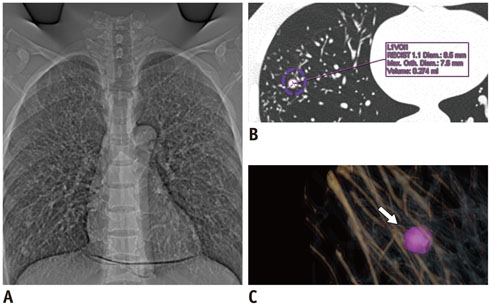
Fig. 1 Chest phantom and volumetric evaluation of synthetic nodule using three-dimensional (3D) volumetry software. Topography (A) for chest dual-source CT examination shows chest phantom and includes vessel structures and synthetic nodules. LungCARE software provides transverse image (B) and volume-rendered image (C) of nodule and 3D volumetric measurement based on pink-color coded volume of interest (arrow).
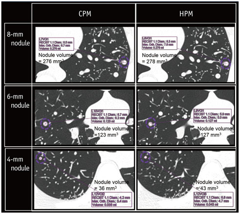
Fig. 2 Volumetric evaluation of synthetic pulmonary nodules (4-mm, 6-mm, and 8-mm in diameter) scanned by dual-source computed tomography in high-pitch mode (HPM) and conventional pitch mode (CPM).
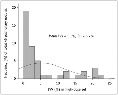
Fig. 3 Histogram of intermode volume variability (IVV) between high-pitch mode and conventional pitch mode for all pulmonary nodules in high-dose set. SD = standard deviation
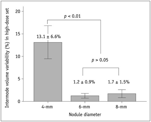
Fig. 4 Comparison of intermode volume variability between high-pitch mode and conventional pitch mode by nodule diameter in high-dose set.
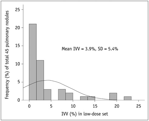
Fig. 5 Histogram of intermode volume variability (IVV) between high-pitch mode and conventional pitch mode for all pulmonary nodules in low-dose set. SD = standard deviation

Fig. 6 Comparison of intermode volume variability between high-pitch mode and conventional pitch mode by nodule diameter in low-dose set.
Reference
-
1. Libby DM, Smith JP, Altorki NK, Pasmantier MW, Yankelevitz D, Henschke CI. Managing the small pulmonary nodule discovered by CT. Chest. 2004; 125:1522–1529.2. Tan BB, Flaherty KR, Kazerooni EA, Iannettoni MD. American College of Chest Physicians. The solitary pulmonary nodule. Chest. 2003; 123:1 Suppl. 89S–96S.3. Diederich S, Hansen J, Wormanns D. Resolving small pulmonary nodules: CT features. Eur Radiol. 2005; 15:2064–2069.4. Nathan MH, Collins VP, Adams RA. Differentiation of benign and malignant pulmonary nodules by growth rate. Radiology. 1962; 79:221–232.5. Weiss W. Peripheral measurable bronchogenic carcinoma. Growth rate and period of risk after therapy. Am Rev Respir Dis. 1971; 103:198–208.6. Kostis WJ, Reeves AP, Yankelevitz DF, Henschke CI. Three-dimensional segmentation and growth-rate estimation of small pulmonary nodules in helical CT images. IEEE Trans Med Imaging. 2003; 22:1259–1274.7. Marten K, Auer F, Schmidt S, Kohl G, Rummeny EJ, Engelke C. Inadequacy of manual measurements compared to automated CT volumetry in assessment of treatment response of pulmonary metastases using RECIST criteria. Eur Radiol. 2006; 16:781–790.8. Lell M, Hinkmann F, Anders K, Deak P, Kalender WA, Uder M, et al. High-pitch electrocardiogram-triggered computed tomography of the chest: initial results. Invest Radiol. 2009; 44:728–733.9. Baumueller S, Alkadhi H, Stolzmann P, Frauenfelder T, Goetti R, Schertler T, et al. Computed tomography of the lung in the high-pitch mode: is breath holding still required? Invest Radiol. 2011; 46:240–245.10. Das M, Ley-Zaporozhan J, Gietema HA, Czech A, Mühlenbruch G, Mahnken AH, et al. Accuracy of automated volumetry of pulmonary nodules across different multislice CT scanners. Eur Radiol. 2007; 17:1979–1984.11. Das M, Mühlenbruch G, Katoh M, Bakai A, Salganicoff M, Stanzel S, et al. Automated volumetry of solid pulmonary nodules in a phantom: accuracy across different CT scanner technologies. Invest Radiol. 2007; 42:297–302.12. Szucs-Farkas Z, Kurmann L, Strautz T, Patak MA, Vock P, Schindera ST. Patient exposure and image quality of low-dose pulmonary computed tomography angiography: comparison of 100- and 80-kVp protocols. Invest Radiol. 2008; 43:871–876.13. Coenen A, Honda O, van der Jagt EJ, Tomiyama N. Computer-assisted solid lung nodule 3D volumetry on CT: influence of scan mode and iterative reconstruction: a CT phantom study. Jpn J Radiol. 2013; 31:677–684.14. Wormanns D, Kohl G, Klotz E, Marheine A, Beyer F, Heindel W, et al. Volumetric measurements of pulmonary nodules at multi-row detector CT: in vivo reproducibility. Eur Radiol. 2004; 14:86–92.15. Funaki A, Ohkubo M, Wada S, Murao K, Matsumoto T, Niizuma S. Application of CT-PSF-based computer-simulated lung nodules for evaluating the accuracy of computer-aided volumetry. Radiol Phys Technol. 2012; 5:166–171.16. Willemink MJ, Leiner T, Budde RP, de Kort FP, Vliegenthart R, van Ooijen PM, et al. Systematic error in lung nodule volumetry: effect of iterative reconstruction versus filtered back projection at different CT parameters. AJR Am J Roentgenol. 2012; 199:1241–1246.17. Wiemker R, Rogalla P, Blaffert T, Sifri D, Hay O, Shah E, et al. Aspects of computer-aided detection (CAD) and volumetry of pulmonary nodules using multislice CT. Br J Radiol. 2005; 78 Spec No 1:S46–S56.18. Christe A, Torrente JC, Lin M, Yen A, Hallett R, Roychoudhury K, et al. CT screening and follow-up of lung nodules: effects of tube current-time setting and nodule size and density on detectability and of tube current-time setting on apparent size. AJR Am J Roentgenol. 2011; 197:623–630.
- Full Text Links
-
- Actions
-
Cited
- CITED
-
- Close
- Share
-
- Similar articles
-
- A study of Parameters in Spiral CT Volumetry Using Balloon Phantoms
- The Difference of Nodule Detection Rates in the Liver According to the Pitch and Slice Thickness in Spiral CT: Experimental Study by Using Artificial Liver Phantom
- Evaluation of Computer Aided Volumetry for Simulated Small Pulmonary Nodules on Computed Tomography
- A Comparison of Two Commercial Volumetry Software Programs in the Analysis of Pulmonary Ground-Glass Nodules: Segmentation Capability and Measurement Accuracy
- Image Quality and Radiation Dose of High-Pitch Dual-Source Spiral Cardiothoracic Computed Tomography in Young Children with Congenital Heart Disease: Comparison of Non-Electrocardiography Synchronization and Prospective Electrocardiography Triggering
