T2 Values of Femoral Cartilage of the Knee Joint: Comparison between Pre-Contrast and Post-Contrast Images
- Affiliations
-
- 1Department of Radiology, Samsung Medical Center, Sungkyunkwan University School of Medicine, Seoul 135-710, Korea. youngcheol.yoon@gmail.com
- 2Department of Preventive Medicine, Kyung Hee University School of Medicine, Seoul 130-872, Korea.
- KMID: 1711486
- DOI: http://doi.org/10.3348/kjr.2014.15.1.123
Abstract
OBJECTIVE
To retrospectively evaluate the relationship between T2 values of pre- and post-contrast magnetic resonance (MR) images of femoral cartilage in patients with varying degrees of osteoarthritis.
MATERIALS AND METHODS
A total of 19 patients underwent delayed gadolinium-enhanced MRI of cartilage. Six regions of interest for T2 value measurement were obtained from pre- and post-contrast T2-weighted, sagittal, multi-slice, multi-echo, source images in each subject. Regions with modified Noyes classification grade 2B and 3 were excluded. Comparison of T2 values between pre- and post-contrast images and T2 values among regions with the grade 0, 1 and 2A groups were statistically analyzed.
RESULTS
Of a total of 114 regions, 79 regions showing grade 0 (n = 46), 1 (n = 18), or 2A (n = 15) were analyzed. The overall and individual T2 values of post-contrast images were significantly lower than those of pre-contrast images (overall, 35.3 +/- 9.2 [mean +/- SD] vs. 29.9 +/- 8.2, p < 0.01; range of individual, 28.9-37.6 vs. 27.1-36.4, p < 0.01). Pearson correlation coefficients showed a strong positive correlation between pre- and post-contrast images (rho-Pearson = 0.712-0.905). T2 values of pre- and post-contrast images of the grade 0 group were significantly lower than those of the grade 1/2A group (pre T2, p = 0.003; post T2, p = 0.006).
CONCLUSION
T2 values of the femoral cartilage of the knee joint are significantly lower on post-contrast images than on pre-contrast images. Furthermore, these T2 values have a strong positive correlation between pre- and post-contrast images.
MeSH Terms
Figure
-

Fig. 1 Example of T2 measurements on map of T2 relaxation time in 51-year-old male patient with osteoarthritis of patellofemoral joint. Three ROIs drawn on femoral cartilage on sagittal, multi-slice, multi-echo, T2-weighted source images (A) were automatically matched to T2 map (B). Mean pixel values of corresponding ROIs on T2 map were automatically measured (numbers in B). T2 values of post-contrast images were measured in same manner. e.g. 4:300 in (A), number on left designates ROI number, and number on right designates mean value of signal intensity in ROI. ROI = regions of interest
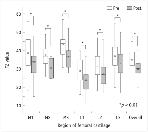
Fig. 2 Boxplots for comparison of T2 values between pre- and post-contrast images. T2 values of all six regions were lower on post-contrast images than on pre-contrast images.
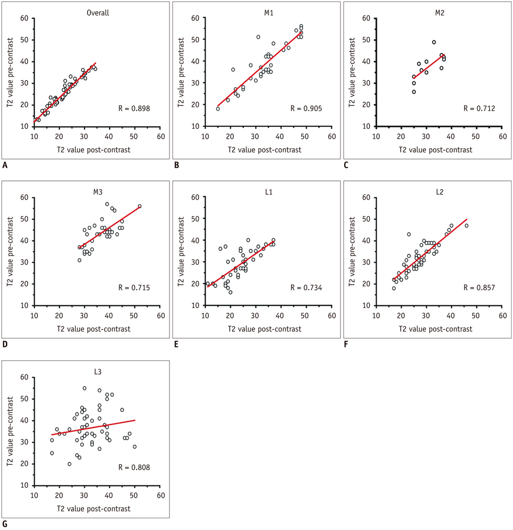
Fig. 3 Scatterplots for correlations of T2 values between pre- and post-contrast images. Pearson correlation coefficients showed strong positive correlation between pre- and post-contrast images overall (A), and in each region (B-G). R = rho-Pearson
Reference
-
1. Nieminen MT, Töyräs J, Laasanen MS, Silvennoinen J, Helminen HJ, Jurvelin JS. Prediction of biomechanical properties of articular cartilage with quantitative magnetic resonance imaging. J Biomech. 2004; 37:321–328.2. Nissi MJ, Töyräs J, Laasanen MS, Rieppo J, Saarakkala S, Lappalainen R, et al. Proteoglycan and collagen sensitive MRI evaluation of normal and degenerated articular cartilage. J Orthop Res. 2004; 22:557–564.3. Chen CT, Fishbein KW, Torzilli PA, Hilger A, Spencer RG, Horton WE Jr. Matrix fixed-charge density as determined by magnetic resonance microscopy of bioreactor-derived hyaline cartilage correlates with biochemical and biomechanical properties. Arthritis Rheum. 2003; 48:1047–1056.4. Wayne JS, Kraft KA, Shields KJ, Yin C, Owen JR, Disler DG. MR imaging of normal and matrix-depleted cartilage: correlation with biomechanical function and biochemical composition. Radiology. 2003; 228:493–499.5. Kurkijärvi JE, Nissi MJ, Kiviranta I, Jurvelin JS, Nieminen MT. Delayed gadolinium-enhanced MRI of cartilage (dGEMRIC) and T2 characteristics of human knee articular cartilage: topographical variation and relationships to mechanical properties. Magn Reson Med. 2004; 52:41–46.6. Bashir A, Gray ML, Burstein D. Gd-DTPA2- as a measure of cartilage degradation. Magn Reson Med. 1996; 36:665–673.7. Bashir A, Gray ML, Hartke J, Burstein D. Nondestructive imaging of human cartilage glycosaminoglycan concentration by MRI. Magn Reson Med. 1999; 41:857–865.8. Nieminen MT, Rieppo J, Silvennoinen J, Töyräs J, Hakumäki JM, Hyttinen MM, et al. Spatial assessment of articular cartilage proteoglycans with Gd-DTPA-enhanced T1 imaging. Magn Reson Med. 2002; 48:640–648.9. Xia Y, Moody JB, Burton-Wurster N, Lust G. Quantitative in situ correlation between microscopic MRI and polarized light microscopy studies of articular cartilage. Osteoarthritis Cartilage. 2001; 9:393–406.10. Gründer W, Wagner M, Werner A. MR-microscopic visualization of anisotropic internal cartilage structures using the magic angle technique. Magn Reson Med. 1998; 39:376–382.11. Nieminen MT, Rieppo J, Töyräs J, Hakumäki JM, Silvennoinen J, Hyttinen MM, et al. T2 relaxation reveals spatial collagen architecture in articular cartilage: a comparative quantitative MRI and polarized light microscopic study. Magn Reson Med. 2001; 46:487–493.12. Kurkijärvi JE, Nissi MJ, Rieppo J, Töyräs J, Kiviranta I, Nieminen MT, et al. The zonal architecture of human articular cartilage described by T2 relaxation time in the presence of Gd-DTPA2-. Magn Reson Imaging. 2008; 26:602–607.13. Nieminen MT, Menezes NM, Williams A, Burstein D. T2 of articular cartilage in the presence of Gd-DTPA2-. Magn Reson Med. 2004; 51:1147–1152.14. Burstein D, Velyvis J, Scott KT, Stock KW, Kim YJ, Jaramillo D, et al. Protocol issues for delayed Gd(DTPA)(2-)-enhanced MRI (dGEMRIC) for clinical evaluation of articular cartilage. Magn Reson Med. 2001; 45:36–41.15. Williams A, Gillis A, McKenzie C, Po B, Sharma L, Micheli L, et al. Glycosaminoglycan distribution in cartilage as determined by delayed gadolinium-enhanced MRI of cartilage (dGEMRIC): potential clinical applications. AJR Am J Roentgenol. 2004; 182:167–172.16. Peterfy CG, Schneider E, Nevitt M. The osteoarthritis initiative: report on the design rationale for the magnetic resonance imaging protocol for the knee. Osteoarthritis Cartilage. 2008; 16:1433–1441.17. Burstein D, Gray M, Mosher T, Dardzinski B. Measures of molecular composition and structure in osteoarthritis. Radiol Clin North Am. 2009; 47:675–686.18. Bredella MA, Tirman PF, Peterfy CG, Zarlingo M, Feller JF, Bost FW, et al. Accuracy of T2-weighted fast spin-echo MR imaging with fat saturation in detecting cartilage defects in the knee: comparison with arthroscopy in 130 patients. AJR Am J Roentgenol. 1999; 172:1073–1080.19. Sonin AH, Pensy RA, Mulligan ME, Hatem S. Grading articular cartilage of the knee using fast spin-echo proton density-weighted MR imaging without fat suppression. AJR Am J Roentgenol. 2002; 179:1159–1166.20. Gillis A, Gray M, Burstein D. Relaxivity and diffusion of gadolinium agents in cartilage. Magn Reson Med. 2002; 48:1068–1071.21. Pedersen M, Mørkenborg J, Jensen FT, Stødkilde-Jørgensen H, Djurhuus JC, Frokiaer J. In vivo measurements of relaxivities in the rat kidney cortex. J Magn Reson Imaging. 2000; 12:289–296.22. Mosher TJ, Smith H, Dardzinski BJ, Schmithorst VJ, Smith MB. MR imaging and T2 mapping of femoral cartilage: in vivo determination of the magic angle effect. AJR Am J Roentgenol. 2001; 177:665–669.23. Dunn TC, Lu Y, Jin H, Ries MD, Majumdar S. T2 relaxation time of cartilage at MR imaging: comparison with severity of knee osteoarthritis. Radiology. 2004; 232:592–598.24. Goodwin DW, Dunn JF. High-resolution magnetic resonance imaging of articular cartilage: correlation with histology and pathology. Top Magn Reson Imaging. 1998; 9:337–347.
- Full Text Links
-
- Actions
-
Cited
- CITED
-
- Close
- Share
-
- Similar articles
-
- MR T2 Map Technique: How to Assess Changes in Cartilage of Patients with Osteoarthritis of the Knee
- A radiological study of the patella and the cartilage of patella by computed tomography following double-contrast arthrography
- Avascular Necrosis of Femoral Head: Findings of Contrast-Enhanced MR Imaging
- Abbreviated Breast Magnetic Resonance Imaging: Background, Evidence From Studies, and Future Considerations
- Usefulness of Postcontrast T2-Weighted Images in Shortening the Total Scan Time of a Gadoxectic Acid Enhanced MRI of the Liver: a Comparison between Precontrast and Postcontrast T2-Weighted Images
