J Korean Soc Radiol.
2010 Mar;62(3):249-256. 10.3348/jksr.2010.62.3.249.
Usefulness of Postcontrast T2-Weighted Images in Shortening the Total Scan Time of a Gadoxectic Acid Enhanced MRI of the Liver: a Comparison between Precontrast and Postcontrast T2-Weighted Images
- Affiliations
-
- 1Department of Radiology, Korea University Guro Hospital, Korea University College of Medicine, Korea. chlee86@hanmail.net
- KMID: 2208920
- DOI: http://doi.org/10.3348/jksr.2010.62.3.249
Abstract
- PURPOSE
We evaluated the effects of gadolinium ethoxybenzyl diethylenetriaminepentaacetic acid (Gd-EOB-DTPA) on the overall image quality and lesion contrast of T2-weighted images (T2WI).
MATERIALS AND METHODS
We reviewed T2WI before and after Gd-EOB-DTPA enhancement in 50 patients with 41 focal liver lesions. Pre- and post-contrast T2WI were analyzed qualitatively and quantitatively. For the qualitative analysis, two abdominal radiologists, blinded to other findings in the study, retrospectively scored the image quality, lesion conspicuity, and artifacts. The quantitative analysis involved the measurement of the liver signal-to-noise ratio (SNR) and the lesion-to-liver contrast-to-noise ratio (CNR).
RESULTS
Image quality and lesion conspicuity were not significantly different among the respiratory-triggered T2-weighted turbo spin-echo sequence (T2TSE), Half-fourier acquisition single-shot turbo spin-echo (HASTE) sequence, and breath-hold T2*- weighted gradient recalled-echo (GRE) sequences. However, there was a trend toward improved lesion conspicuity in post-contrast T2TSE. The quantitative analysis indicated that the SNR for post-contrast images was significantly higher than for pre-contrast images on T2TSE (p=0.04). No significant differences were seen in the CNR of focal lesions, but there was a trend towards an increase in the CNR for postcontrast T2WI of hepatocellular carcinoma.
CONCLUSION
Compared to pre-contrast T2WI, post-contrast T2WI showed no handicap in the overall image quality and lesion contrast. Therefore, to shorten the total scan time during Gd-EOB-DTPA enhanced MRI, pre-contrast T2WI can be replaced by post-contrast T2WI.
MeSH Terms
Figure
-
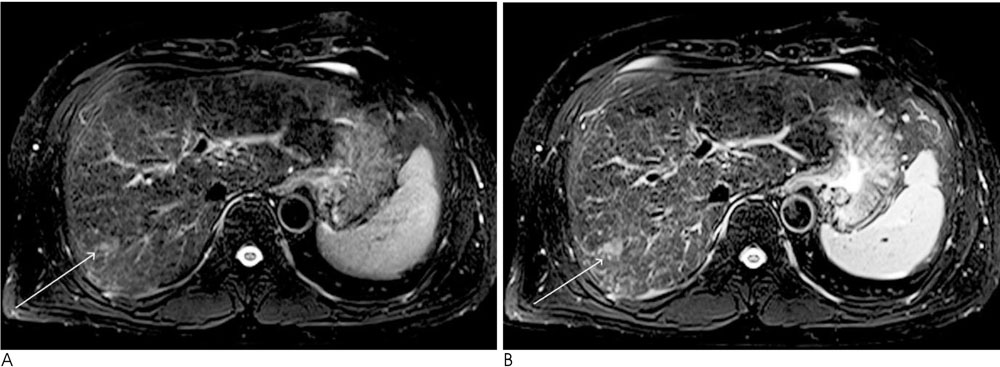
Fig. 1 50-year-old man with hepatocellular carcinoma (arrow). A, B. respiratory triggered T2TSE MR images before (A) and after (B) the administration of Gd-EOB-DTPA. T2TSE MR images after the administration of Gd-EOB-DTPA was rated as having better lesion conspicuity.
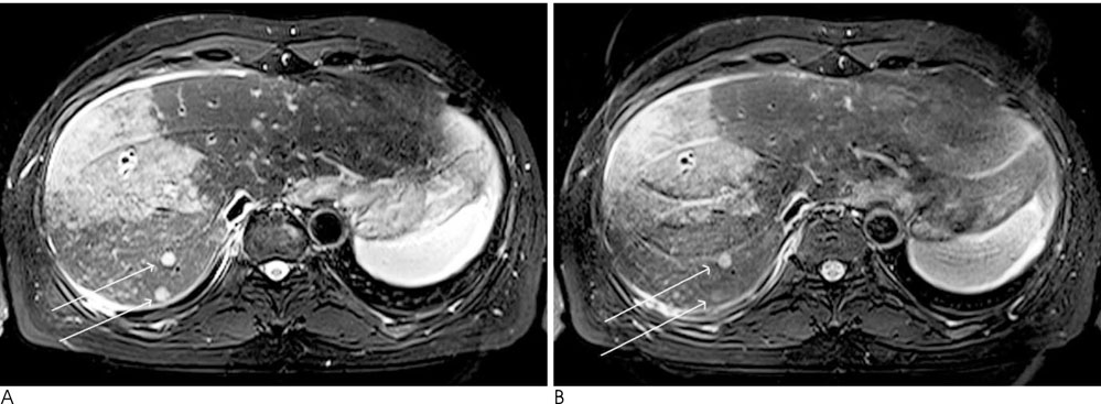
Fig. 2 A 66-year-old man with cholangiocarcinoma and intrahepatic metastasis (arrows). A, B. respiratory triggered T2TSE MR images before (A) and after (B) the administration of Gd-EOB-DTPA. T2TSE MR images before the administration of Gd-EOB-DTPA was rated as having better lesion conspicuity.
Reference
-
1. Weinmann HJ, Schuhmann-Giampieri G, Schmitt-Willich H, Vogler H, Frenzel T, Gries H. A new lipophilic gadolinium chelate as a tissue-specific contrast medium for MRI. Magn Reson Med. 1991; 22:233–237. 2422. Hammerstingl R, Huppertz A, Breuer J, Balzer T, Blakeborough A, Carter R, et al. Diagnostic efficacy of gadoxetic acid (Primovist)-enhanced MRI and spiral CT for a therapeutic strategy: comparison with intraoperative and histopathologic findings in focal liver lesions. Eur Radiol. 2008; 18:457–467.3. Halavaara J, Breuer J, Ayuso C, Balzer T, Bellin MF, Blomqvist L, et al. Liver tumor characterization: comparison between liver-specific gadoxetic acid disodium-enhanced MRI and biphasic CT—a multicenter trial. J Comput Assist Tomogr. 2006; 30:345–354.4. Huppertz A, Balzer T, Blakeborough A, Breuer J, Giovagnoni A, Heinz-Peer G, et al. Improved detection of focal liver lesions at MR imaging: multicenter comparison of gadoxetic acid-enhanced MR images with intraoperative findings. Radiology. 2004; 230:266–275.5. Zizka J, Klzo L, Ferda J, Mrklovsky M, Bukac J. Dynamic and delayed contrast enhancement in upper abdominal MRI studies: comparison of gadoxetic acid and gadobutrol. Eur J Radiol. 2007; 62:186–191.6. Runge VM. A comparison of two MR hepatobiliary gadolinium chelates: Gd-BOPTA and Gd-EOB-DTPA. J Comput Assist Tomogr. 1998; 22:643–650.7. Tanimoto A, Satoh Y, Yuasa Y, Jinzaki M, Hiramatsu K. Performance of Gd-EOB-DTPA and superparamagnetic iron oxide particles in the detection of primary liver cancer: a comparative study by alternative free-response receiver operating characteristic analysis. J Magn Reson Imaging. 1997; 7:120–124.8. Vogl TJ, Kummel S, Hammerstingl R, Schellenbeck M, Schumacher G, Balzer T, et al. Liver tumors: comparison of MR imaging with Gd-EOB-DTPA and Gd-DTPA. Radiology. 1996; 200:59–67.9. Reimer P, Schneider G, Schima W. Hepatobiliary contrast agents for contrast-enhanced MRI of the liver: properties, clinical development and applications. Eur Radiol. 2004; 14:559–578.10. Huppertz A, Haraida S, Kraus A, Zech CJ, Scheidler J, Breuer J, et al. Enhancement of focal liver lesions at gadoxetic acid-enhanced MR imaging: correlation with histopathologic findings and spiral CT—initial observations. Radiology. 2005; 234:468–478.11. Ohtomo K, Itai Y, Yoshikawa K, Kokubo T, Iio M. Hepatocellular carcinoma and cavernous hemangioma: differentiation with MR imaging. Efficacy of T2 values at 0.35 and 1.5 T. Radiology. 1988; 168:621–623.12. Chang SD, Thoeni RF. Effect of T1 shortening on T2-weighted MRI sequences: comparison of hepatic mass conspicuity on images acquired before and after gadolinium enhancement. AJR Am J Roentgenol. 2008; 190:1318–1323.13. Jeong YY, Mitchell DG, Holland GA. Liver lesion conspicuity: T2-weighted breath-hold fast spin-echo MR imaging before and after gadolinium enhancement—initial experience. Radiology. 2001; 219:455–460.14. Zech CJ, Herrmann KA, Reiser MF, Schoenberg SO. MR imaging in patients with suspected liver metastases: value of liver-specific contrast agent Gd-EOB-DTPA. Magn Reson Med Sci. 2007; 6:43–52.15. Helmberger T, Gregor M, Holzknecht N, Gauger J, Rau H, Reiser MF. Comparison of dual-phase helical CT with native and ferum oxide-enhanced magnetic resonance imaging in detection and characterization of focal liver lesions. Radiologe. 1999; 39:678–684.16. Schuhmann-Giampieri G, Schmitt-Willich H, Press WR, Negishi C, Weinmann HJ, Speck U. Preclinical evaluation of Gd-EOB-DTPA as a contrast agent in MR imaging of the hepatobiliary system. Radiology. 1992; 183:59–64.17. Stroszczynski C, Gaffke G, Gnauck M, Streitparth F, Wieners G, Lopez-Haninnen E. Current status of MRI diagnostics with liverspecific contrast agents. Gd-EOB-DTPA and Gd-BOPTA. Radiologe. 2004; 44:1185–1191.18. Outwater E, Schnall MD, Braitman LE, Dinsmore BJ, Kressel HY. Magnetization transfer of hepatic lesions: evaluation of a novel contrast technique in the abdomen. Radiology. 1992; 182:535–540.19. Reimer P, Rummeny EJ, Daldrup HE, Hesse T, Balzer T, Tombach B, et al. Enhancement characteristics of liver metastases, hepatocellular carcinomas, and hemangiomas with Gd-EOB-DTPA: preliminary results with dynamic MR imaging. Eur Radiol. 1997; 7:275–280.
- Full Text Links
-
- Actions
-
Cited
- CITED
-
- Close
- Share
-
- Similar articles
-
- Hemangioma and Hepatocellular Carcinoma: Distinction with Superparamagnetic Iron Oxide-Enhanced MR Imaging
- Comparative Study on the Size of Hepatic Metastases Between Pre- and Postcontrast CT
- Ring Lesions in MR Imaging of the Liver
- Adenomyosis of the Uterus: Recognition of a Characteristic Enhancement Pattern on Contrast-enhanced Dynamic MRI
- MR Imaging findings of Diffuse Axonal Injury: Comparison of T2*-weighted Gradient Images and T1- andT2-weighted Spin-Echo Images
