Interpretation of Digital Chest Radiographs: Comparison of Light Emitting Diode versus Cold Cathode Fluorescent Lamp Backlit Monitors
- Affiliations
-
- 1Department of Radiology and Center for Imaging Science, Samsung Medical Center, Sungkyunkwan University School of Medicine, Seoul 135-710, Korea. mjchung@skku.edu
- KMID: 1711466
- DOI: http://doi.org/10.3348/kjr.2013.14.6.968
Abstract
OBJECTIVE
To compare the diagnostic performance of light emitting diode (LED) backlight monitors and cold cathode fluorescent lamp (CCFL) monitors for the interpretation of digital chest radiographs.
MATERIALS AND METHODS
We selected 130 chest radiographs from health screening patients. The soft copy image data were randomly sorted and displayed on a 3.5 M LED (2560 x 1440 pixels) monitor and a 3 M CCFL (2048 x 1536 pixels) monitor. Eight radiologists rated their confidence in detecting nodules and abnormal interstitial lung markings (ILD). Low dose chest CT images were used as a reference standard. The performance of the monitor systems was assessed by analyzing 2080 observations and comparing them by multi-reader, multi-case receiver operating characteristic analysis. The observers reported visual fatigue and a sense of heat. Radiant heat and brightness of the monitors were measured.
RESULTS
Measured brightness was 291 cd/m2 for the LED and 354 cd/m2 for the CCFL monitor. Area under curves for nodule detection were 0.721 +/- 0.072 and 0.764 +/- 0.098 for LED and CCFL (p = 0.173), whereas those for ILD were 0.871 +/- 0.073 and 0.844 +/- 0.068 (p = 0.145), respectively. There were no significant differences in interpretation time (p = 0.446) or fatigue score (p = 0.102) between the two monitors. Sense of heat was lower for the LED monitor (p = 0.024). The temperature elevation was 6.7degrees C for LED and 12.4degrees C for the CCFL monitor.
CONCLUSION
Although the LED monitor had lower maximum brightness compared with the CCFL monitor, soft copy reading of the digital chest radiographs on LED and CCFL showed no difference in terms of diagnostic performance. In addition, LED emitted less heat.
Keyword
MeSH Terms
Figure
-
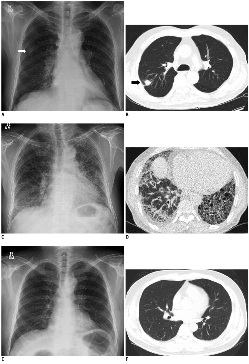
Fig. 1 Examples of selected chest radiographs and reference CT images. Those with nodue (A, B), increased interstitial markings (C, D) and neither abnormality (E, F), respectively.
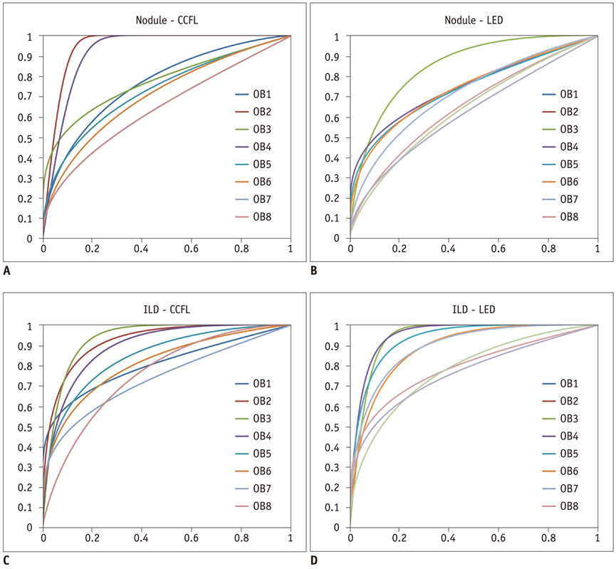
Fig. 2 ROC curves for nodules (A, B) and ILD (C, D). X-axis value = false positive rate (1-specificity), Y-axis value = true positive rate (sensitivity), ROC = receiver operating characteristic, ILD = interstitial lung marking, CCFL = cold cathode fluorescent lamp, LED = light emitting diode
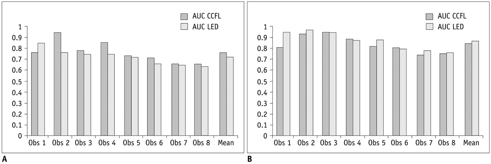
Fig. 3 Individual and mean ROC and AUC values for nodules (A) and ILD (B) using CCFL and LED monitor displays. Observers 1, 2, 3, and 4 were board certified radiologists and observers 5, 6, 7, and 8 were residents. AUC = area under curve, ROC = receiver operating characteristic, ILD = interstitial lung marking, CCFL = cold cathode fluorescent lamp, LED = light emitting diode
Reference
-
1. Iwano S, Ishigaki T, Shimamoto K, Inamura K, Maeda T, Ikeda M, et al. Detection of subtle pulmonary disease on CR chest images: monochromatic CRT monitor vs color CRT monitor. Eur Radiol. 2001; 11:59–64.2. American college of Radiology (ACR) Web site. ACR-AAPM-SIIM Technical standard for electronic practice of medical imaging. revised 2012. Accessed January 1, 2013. http://www.acr.org/.3. Muraoka T, Nakashima N, Mizushina S, Ikeda H, Shimodaira Y. Subjective Evaluation of Physiological Fatigue in Video Data Terminal Operation. In : Proceedings of Image Processing, Image Quality, Image Capture, System Conference, The Society for Imaging Science and Technology; Portland. 1998. p. 266–270.4. Zwanenburg M, Dunn T, Stich A, Schwelder W, Plotz L. High-Efficiency LEDs for LCD Backlight. SID Symp Dig Tech Pap. 2004; 1222–1225. DOI:10.1889/1.1821338.5. Anandan M. LED Backlight: Enhancement of picture quality on LCD screen. Proc of ASID. 2006; 130–134.6. Dorfman DD, Berbaum KS, Metz CE. Receiver operating characteristic rating analysis. Generalization to the population of readers and patients with the jackknife method. Invest Radiol. 1992; 27:723–731.7. Obuchowski NA. New methodological tools for multiple-reader ROC studies. Radiology. 2007; 243:10–12.8. Indrajit I, Verma B. Monitor displays in radiology: Part 1. Indian J Radiol Imaging. 2009; 19:24–28.9. Usami H, Ikeda M, Ishigaki T, Fukushima H, Shimamoto K. The influence of liquid crystal display (LCD) monitors on observer performance for the detection of nodular lesions on chest radiographs. Eur Radiol. 2006; 16:726–732.10. Sund P, Båth M, Kheddache S, Månsson LG. Comparison of visual grading analysis and determination of detective quantum efficiency for evaluating system performance in digital chest radiography. Eur Radiol. 2004; 14:48–58.11. Understanding LCD Monitors: Brightness and Contrast. Accessed January 3, 2013. http://www.nettivire.fi/kirkkaustiedote.pdf/.12. Carrein G. Barco Web site. Barco White Paper: 10 reasons to use a medical display system. published 2006. Accessed January 5, 2013. http://www.barco.com/barcoview/downloads/10_reasons_to_use_a_medical_display_system.pdf.13. American college of Radiology (ACR) Web site. ACR standard for digital image data management. amended 2006. Accessed January 2, 2013. http://www.acr.org/.14. Goo JM, Choi JY, Im JG, Lee HJ, Chung MJ, Han D, et al. Effect of monitor luminance and ambient light on observer performance in soft-copy reading of digital chest radiographs. Radiology. 2004; 232:762–766.15. Krupinski EA. Medical grade vs off-the-shelf color displays: influence on observer performance and visual search. J Digit Imaging. 2009; 22:363–368.
- Full Text Links
-
- Actions
-
Cited
- CITED
-
- Close
- Share
-
- Similar articles
-
- Liquid-Crystal Display Monitors and Cathode-Ray Tube Monitors: A Comparison of Observer Performance in the Detection of Small Solitary Pulmonary Nodules
- The shear bond strength and adhesive failure pattern in bracket bonding with different light-curing methods
- Topical Photodynamic Therapy for Treatment of Actinic Keratosis Using Light-Emitting Diode (LED) Device
- Transfer printing based manufacturing process for flexible micro-light emitting diode phototherapy devices
- Comparison of Contrast Sensitivity and Color Vision according to the Different Illumination in Patients with Cataract
