Are Spinal or Paraspinal Anatomic Markers Helpful for Vertebral Numbering and Diagnosing Lumbosacral Transitional Vertebrae?
- Affiliations
-
- 1Department of Radiology, Gazi University School of Medicine, Ankara 06510, Turkey. niltokgoz@yahoo.com
- KMID: 1705582
- DOI: http://doi.org/10.3348/kjr.2014.15.2.258
Abstract
OBJECTIVE
To evaluate the value of spinal and paraspinal anatomic markers in both the diagnosis of lumbosacral transitional vertebrae (LSTVs) and identification of vertebral levels on lumbar MRI.
MATERIALS AND METHODS
Lumbar MRI from 1049 adult patients were studied. By comparing with the whole-spine localizer, the diagnostic errors in numbering vertebral segments on lumbar MRI were evaluated. The morphology of S1-2 disc, L5 and S1 body, and lumbar spinous processes (SPs) were evaluated by using sagittal MRI. The positions of right renal artery (RRA), superior mesenteric artery, aortic bifurcation (AB) and conus medullaris (CM) were described.
RESULTS
The diagnostic error for evaluation of vertebral segmentation on lumbar MRI alone was 14.1%. In lumbarization, all patients revealed a well-formed S1-2 disc with squared S1 body. A rhombus-shaped L5 body in sacralization and a rectangular-shaped S1 body in lumbarization were found. The L3 had the longest SP. The most common sites of spinal and paraspinal structures were: RRA at L1 body (53.6%) and L1-2 disc (34.1%), superior mesenteric artery at L1 body (55.1%) and T12-L1 disc (31.6%), and AB at L4 body (71.1%). CM had variable locations, changing from the T12-L1 disc to L2 body. They were located at higher sacralization and lower lumbarization.
CONCLUSION
The spinal morphologic features and locations of the spinal and paraspinal structures on lumbar MRI are not completely reliable for the diagnosis of LSTVs and identification on the vertebral levels.
Keyword
MeSH Terms
-
Adolescent
Adult
Aged
Aged, 80 and over
Anatomic Landmarks/*anatomy & histology
Aorta, Abdominal/anatomy & histology
Diagnostic Errors
Female
Humans
Intervertebral Disc/anatomy & histology
Lumbar Vertebrae/*anatomy & histology
Lumbosacral Region
Magnetic Resonance Imaging
Male
Mesenteric Artery, Superior/anatomy & histology
Middle Aged
Renal Artery/anatomy & histology
Reproducibility of Results
Sacrum/*anatomy & histology
Spinal Cord/anatomy & histology
Spine
Young Adult
Figure
-

Fig. 1 Example of sagittal whole-spine localizer image with half-Fourier acquisition single shot turbo spin-echo sequence for numbering lumbar vertebrae.
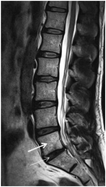
Fig. 2 Sagittal T2-weighted MRI of man with lumbarization of S1 vertebra (thick arrow). There is well-formed S1-2 disc (thin arrow) with squaring of S1 vertebral body (thick arrow).

Fig. 3 Sagittal T2-weighted MRI of woman demonstrates sacralization of L5 vertebra (arrow). L5 body shows rhombus shape similar to S1 vertebra.

Fig. 4 Sagittal T2-weighted images (A-C) of man with normal lumbar segmentation. A. Proximal right renal artery (arrow) is located at L1-2 disc space. B. Root of superior mesenteric artery (arrow) is positioned at L1 body. C. Aortic bifurcation (arrow) is located at L4 body.
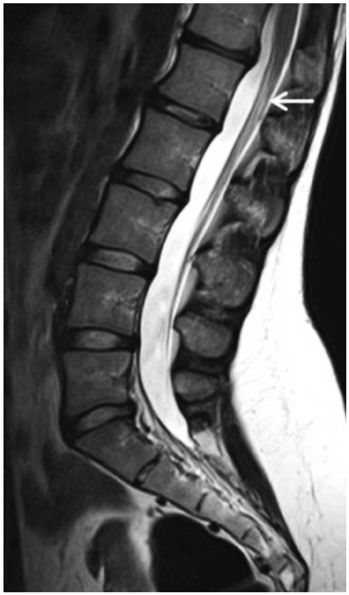
Fig. 5 Sagittal T2-weighted MRI of woman with normal lumbar segmentation shows that conus medullaris (arrow) is located at L1 body.
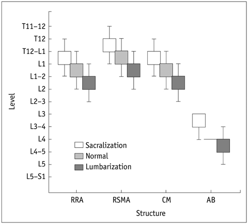
Fig. 6 Comparison on locational distributions of spinal and paraspinal structures in study groups. RRA = right renal artery, RSMA = root of superior mesenteric artery, CM = conus medullaris, AB = aortic bifurcation
Reference
-
1. Hughes RJ, Saifuddin A. Numbering of lumbosacral transitional vertebrae on MRI: role of the iliolumbar ligaments. AJR Am J Roentgenol. 2006; 187:W59–W65.2. Hughes RJ, Saifuddin A. Imaging of lumbosacral transitional vertebrae. Clin Radiol. 2004; 59:984–991.3. Konin GP, Walz DM. Lumbosacral transitional vertebrae: classification, imaging findings, and clinical relevance. AJNR Am J Neuroradiol. 2010; 31:1778–1786.4. Lee CH, Park CM, Kim KA, Hong SJ, Seol HY, Kim BH, et al. Identification and prediction of transitional vertebrae on imaging studies: anatomical significance of paraspinal structures. Clin Anat. 2007; 20:905–914.5. Carrino JA, Campbell PD Jr, Lin DC, Morrison WB, Schweitzer ME, Flanders AE, et al. Effect of spinal segment variants on numbering vertebral levels at lumbar MR imaging. Radiology. 2011; 259:196–202.6. Lee CH, Seo BK, Choi YC, Shin HJ, Park JH, Jeon HJ, et al. Using MRI to evaluate anatomic significance of aortic bifurcation, right renal artery, and conus medullaris when locating lumbar vertebral segments. AJR Am J Roentgenol. 2004; 182:1295–1300.7. Peh WC, Siu TH, Chan JH. Determining the lumbar vertebral segments on magnetic resonance imaging. Spine (Phila Pa 1976). 1999; 24:1852–1855.8. Castellvi AE, Goldstein LA, Chan DP. Lumbosacral transitional vertebrae and their relationship with lumbar extradural defects. Spine (Phila Pa 1976). 1984; 9:493–495.9. MacGibbon B, Farfan HF. A radiologic survey of various configurations of the lumbar spine. Spine (Phila Pa 1976). 1979; 4:258–266.10. Hahn PY, Strobel JJ, Hahn FJ. Verification of lumbosacral segments on MR images: identification of transitional vertebrae. Radiology. 1992; 182:580–581.11. O'Driscoll CM, Irwin A, Saifuddin A. Variations in morphology of the lumbosacral junction on sagittal MRI: correlation with plain radiography. Skeletal Radiol. 1996; 25:225–230.12. Byun WM, Kim JW, Lee JK. Differentiation between symptomatic and asymptomatic extraforaminal stenosis in lumbosacral transitional vertebra: role of three-dimensional magnetic resonance lumbosacral radiculography. Korean J Radiol. 2012; 13:403–411.13. Bron JL, van Royen BJ, Wuisman PI. The clinical significance of lumbosacral transitional anomalies. Acta Orthop Belg. 2007; 73:687–695.14. Malanga GA, Cooke PM. Segmental anomaly leading to wrong level disc surgery in cauda equina syndrome. Pain Physician. 2004; 7:107–110.15. Wigh RE. The thoracolumbar and lumbosacral transitional junctions. Spine (Phila Pa 1976). 1980; 5:215–222.16. Wigh RE. Phylogeny and the herniated disc. South Med J. 1979; 72:1138–1143.17. Wigh RE, Anthony HF Jr. Transitional lumbosacral discs. probability of herniation. Spine (Phila Pa 1976). 1981; 6:168–171.18. Sobottke R, Koy T, Röllinghoff M, Siewe J, Kreitz T, Müller D, et al. Computed tomography measurements of the lumbar spinous processes and interspinous space. Surg Radiol Anat. 2010; 32:731–738.19. Tan SH, Teo EC, Chua H. Quantitative three-dimensional anatomy of lumbar vertebrae in Singaporean Asians. Eur Spine J. 2002; 11:152–158.20. Ralston MD, Dykes TA, Applebaum BI. Verification of lumbar vertebral bodies. Radiology. 1992; 185:615–616.21. Healy JC, Borley NR, Mundy AR, Collins P, Wigley C. True pelvis, pelvic floor and perineum. In : Standring S, editor. Gray's anatomy: the anatomical basis of clinical practice. 39th ed. New York: Elsevier Churchill Livingstone;2005. p. 1360.22. Chithriki M, Jaibaji M, Steele RD. The anatomical relationship of the aortic bifurcation to the lumbar vertebrae: a MRI study. Surg Radiol Anat. 2002; 24:308–312.23. Kumar S, Neyaz Z, Gupta A. The utility of 64 channel multidetector CT angiography for evaluating the renal vascular anatomy and possible variations: a pictorial essay. Korean J Radiol. 2010; 11:346–354.24. Williams A, Newell RLM, Collins P. Macroscopic anatomy of the spinal cord and spinal nerves. In : Standring S, editor. Gray's anatomy: the anatomical basis of clinical practice. 39th ed. New York: Elsevier Churchill Livingstone;2005. p. 775.
- Full Text Links
-
- Actions
-
Cited
- CITED
-
- Close
- Share
-
- Similar articles
-
- A Case of Bertolloti Syndrome
- Incidence Rate of Lumbosacral Transitional Vertebrae and Measurement of their Cross-sectional Areas of Vertebral Canal and Dural Sac Using Magnetic Resonance Imaging
- The prevalence and clinical significance of transitional vertebrae: a radiologic investigation using whole spine spiral three-dimensional computed tomographic images
- Far-out Stenosis by the Lumbosacral Transitional Vertebrae
- The Role of Transitional Vertebra in Spondylolysis and Spondyloytic Spondylolisthesis
