J Vet Sci.
2013 Sep;14(3):337-343. 10.4142/jvs.2013.14.3.337.
Evaluation of bone healing in canine tibial defects filled with cortical autograft, commercial-DBM, calf fetal DBM, omentum and omentum-calf fetal DBM
- Affiliations
-
- 1Department of Veterinary Surgery and Radiology, School of Veterinary Medicine, Shahrekord University, Shahrekord, Iran. dr.bigham@gmail.com
- 2Department of Veterinary Pathobiology, School of Veterinary Medicine, Shahrekord University, Shahrekord, Iran.
- 3Department of Veterinary Pathobiology, School of Veterinary Medicine, Shiraz University, Shiraz, Iran.
- KMID: 1705565
- DOI: http://doi.org/10.4142/jvs.2013.14.3.337
Abstract
- The present study was conducted to compare the effects of xenogenic bovine fetal demineralized bone matrix (DBM), commercial DBM, omentum, omentum-calf fetal DBM, cortical autograft and xenogenic cartilage powder on the healing of tibial defects in a dog model to determine the best material for bone healing. Seven male adult mongrel dogs, weighing 26.2 +/- 2.5 kg, were used in this study. Seven holes with a diameter of 4-mm were created and then filled with several biomaterials. Radiographs were taken postoperatively on day 1 and weeks 2, 4, 6, 8. The operated tibias were removed on the 56th postoperative day and histopathologically evaluated. On postoperative days 14, 42 and 56, the lesions of the control group were significantly inferior to those in the other group (p < 0.05). On the 28th postoperative day, the autograft group was significantly superior to the control and omentum groups (p < 0.05). Moreover, calf fetal DBM was significantly superior to the control group. There was no significant difference between the histopathological sections of all groups. Overall, the omentum and omentum-DBM groups were superior to the control group, but inferior to the autograft, commercial-DBM, calf fetal DBM and calf fetal cartilage groups.
Keyword
MeSH Terms
Figure
-
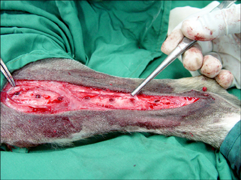
Fig. 1 Seven bone defects were created for implantation of seven different biomaterials in tibial bone.
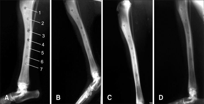
Fig. 2 Radiological evaluation on the 14th (A), 28th (B), 42nd (C) and 58th (D) postoperative days. 1: control, 2: autograft, 3: omentum, 4: omentum-calf fetal demineralized bone matrix (DBM), 5: commercial DBM, 6: calf fetal-DBM, 7: cartilage group.
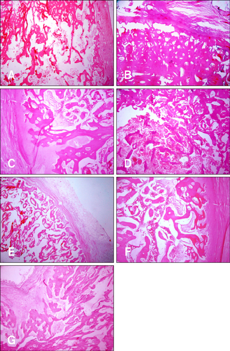
Fig. 3 Histopathological findings showed normal structure of trabecular bone in the defect area in all groups with various phases of remodeling. (A) Control. (B) Autograft. (C) Commercial-DBM. (D) Calf fetal-DBM. (E) Omentum. (F) Omentum-calf fetal DBM. (G) Cartilage group (H&E stain, ×10).
Reference
-
1. Alexander JW. Bone grafting. Vet Clin North Am Small Anim Pract. 1987; 17:811–819.
Article2. Alexander JW. Leonard's Orthopedic Surgery of the Dog and Cat. 3rd ed. Florida: WB Sounders;1985. p. 43–48.3. Arrington ED, Smith WJ, Chambers HG, Bucknell AL, Davino NA. Complications of iliac crest bone graft harvesting. Clin Orthop Relat Res. 1996; 329:300–309.
Article4. Bauer TW, Muschler GF. Bone graft materials: an overview of the basic science. Clin Orthop Relat Res. 2000; 371:10–27.5. Bostrom MPG, Lane JM, Berberian WS, Missri AAE, Tomin E, Weiland A, Doty SB, Glaser D, Rosen VM. Immunolocalization and expression of bone morphogenetic proteins 2 and 4 in fracture healing. J Orthop Res. 1995; 13:357–367.
Article6. Burwell RG. The function of bone marrow in the incorporation of a bone graft. Clin Orthop Relat Res. 1985; 200:125–141.
Article7. Cook SD, Baffes GC, Wolfe MW, Sampath TK, Rueger DC. Recombinant human bone morphogenetic protein-7 induces healing in a canine long-bone segmental defect model. Clin Orthop Relat Res. 1994; 301:302–312.
Article8. Damien CJ, Parsons JR. Bone graft and bone graft substitutes: a review of current technology and applications. J Appl Biomater. 1991; 2:187–208.
Article9. Deckers MML, Karperien M, van der, Yamashita T, Papapoulos SE, Löwik CWGM. Expression of vascular endothelial growth factors and their receptors during osteoblast differentiation. Endocrinology. 2000; 141:1667–1674.
Article10. Diaz-Flores L, Gutierrez R, Lopez-Alonso A, Gonzalez R, Varela H. Pericytes as a supplementary source of osteoblasts in periosteal osteogenesis. Clin Orthop. 1992; 275:280–286.
Article11. Fitch R, Kerwin S, Sinibaldi KR, Newman-Gage H. Bone autografts and allografts in dogs. Compend Contin Educ Pract Vet. 1997; 19:558–575.12. Forell EB, Straw RC, Powers BE, Johnson J, Cooper MF, Withrow J St. Evaluation of the osteoinductive capacity of canine demineralized bone matrix in heterotopic muscle sites of athymic rats. Vet Comp Orthop Traumatol. 1993; 1:25–32.
Article13. Fox SM. Cancellous bone grafting in the dog: an overview. J Am Anim Hosp Assoc. 1984; 20:840–848.14. Friedlaender GE. Bone grafts: the basic science rationale for clinical applications. J Bone Joint Surg Am. 1987; 69:786–790.
Article15. Glowacki J. Angiogenesis in fracture repair. Clin Orthop Relat Res. 1998; 355:Suppl. S82–S89.
Article16. Gordon SD, Warren RF. Autogenous diced cartilage transplants to bone: an experimental study. Ann Surg. 1947; 125:237–240.
Article17. Guizzardi S, Di Silvestre M, Scandroglio R, Ruggeri A, Savini R. Implants of heterologous demineralized bone matrix for induction of posterior spinal fusion in rats. Spine (Phila Pa 1976). 1992; 17:701–707.
Article18. Heiple KG, Goldberg VM, Powell AE, Bos GD, Zika JM. Biology of cancellous bone grafts. Orthop Clin North Am. 1987; 18:179–185.
Article19. Inoue K, Ohgushi H, Yoshikawa T, Okumura M, Sempuku T, Tamai S, Dohi Y. The effect of aging on bone formation in porous hydroxyapatite: biochemical and histological analysis. J Bone Miner Res. 1997; 12:989–994.
Article20. Jin DD. Bone matrix gelatin. Clinical application in 38 cases. Zhonghua Wai Ke Za Zhi. 1991; 29:312–314.21. Khan SN, Cammisa FPJ, Sandhu HS, Diwan AD, Girardi FP, Lane JM. The biology of bone grafting. J Am Acad Orthop Surg. 2005; 13:77–86.
Article22. Kirker-Head AC. Recombinant bone morphogenetic proteins: novel substances for enhancing bone healing. Vet Surg. 1995; 24:408–419.
Article23. Loredo GA, MacDonald MH, Benton HP. Regulation of glycosaminoglycan metabolism by bone morphogenetic protein-2 in equine cartilage explant cultures. Am J Vet Res. 1996; 57:554–559.24. Martin GJ Jr, Boden SD, Titus L, Scarborough NL. New formulations of demineralized bone matrix as a more effective graft alternative in experimental posterolateral lumbar spine arthrodesis. Spine (Phila Pa 1976). 1999; 24:637–645.
Article25. McLaughlin RM, Roush JK. Autogenous cancellous and corticocancellous bone grafting. Vet Med. 1998; 98:1071–1074.26. Moore MAS. Putting the neo into neoangiogenesis. J Clin Invest. 2002; 109:313–315.
Article27. Piermattei DL, Flo GL. Brinker, Piermattei, and Flo's Handbook of Small Animal Orthopedics and Fracture Repair. 3rd ed. Philadelphia: WB Saunders;1997. p. 147–153.28. Reddi AH. Bone morphogenetic proteins, bone marrow stromal cells, and mesenchymal stem cells. Maureen Owen revisited. Clin Orthop Relat Res. 1995; 313:115–119.29. Reddi AH, Huggins C. Biochemical sequences in the transformation of normal fibroblasts in adolescent rats. Proc Natl Acad Sci U S A. 1972; 69:1601–1605.
Article30. Riley EH, Lane JM, Urist MR, Lyons KM, Lieberman JR. Bone morphogenetic protein-2: biology and applications. Clin Orthop Relat Res. 1996; 324:39–46.31. Tanaka T, Fujii K, Ohta M, Soshi S, Kitamura A, Murota K. Use of a guanidine extract of demineralized bone in the treatment of osteochondral defects of articular cartilage. J Orthop Res. 1995; 13:464–469.
Article32. Takada T, Kamei Y, Iwata T, Yokoi T, Torii S. Effect of omental lipid fraction on enhancement of skin flap survival. Ann Plast Surg. 1998; 41:70–74.
Article33. Tuli SM, Singh AD. The osteoinductive property of decalcified bone matrix: an experimental study. J Bone Joint Surg Br. 1978; 60:116–123.34. Urist MR. Bone: formation by autoinduction. Science. 1965; 150:893–899.
Article35. Urist MR, Mikulski AJ, Lietz A. Solubilized and insolubilized bone morphogenetic protein. Proc Natl Acad Sci U S A. 1979; 76:1828–1832.
Article36. Urist MR, Sato K, Brownell AG, Malinin TI, Lietze A, Huo YK, Prolo DJ, Oklund S, Finerman GA, DeLange RJ. Human bone morphogenetic protein (hBMP). Proc Soc Exp Biol Med. 1983; 173:194–199.
Article37. Urist MR, Silverman BF, Büring K, Dubuc FL, Rosenberg JM. The bone induction principle. Clin Orthop Relat Res. 1967; 53:243–283.38. Urist MR, Strates BS. Bone formation in implants of partially and wholly demineralized bone matrix: including observations on acetone-fixed intra and extracellular proteins. Clin Orthop Relat Res. 1970; 71:271–278.39. Vail TB, Trotter GW, Powers BE. Equine demineralized bone matrix: relationship between particle size and osteoinduction. Vet Surg. 1994; 23:386–395.
Article40. Van Heest A, Swiontkowski M. Bone-graft substitutes. Lancet. 1999; 353:Suppl 1. SI28–SI29.
Article
- Full Text Links
-
- Actions
-
Cited
- CITED
-
- Close
- Share
-
- Similar articles
-
- Effect of Murine Adipose Derived Stem Cell(ADSC) on Bone Induction of Demineralized Bone Matrix(DBM) in a Rat Calvarian Defect Model
- Compatibility of Self-setting DBM-CP Composites in Percutaneous Kyphoplasty
- Demineralized Bone Matrix Injection in Consolidation Phase Enhances Bone Regeneration in Distraction Osteogenesis via Endochondral Bone Formation
- Benefits of a Demineralized Bone Matrix in Osteoporotic Intertrochanteric Femoral Fracture Patients
- Comparison of Fusion Rate between Demineralized Bone Matrix versus Autograft in Lumbar Fusion : Meta-Analysis
