Korean J Radiol.
2013 Jun;14(3):520-524. 10.3348/kjr.2013.14.3.520.
Langerhans Cell Sarcoma in Two Young Children: Imaging Findings on Initial Presentation and Recurrence
- Affiliations
-
- 1Department of Radiology, Seoul St. Mary's Hospital, College of Medicine, The Catholic University of Korea, Seoul 137-701, Korea. saim@catholic.ac.kr
- 2Department of Pediatrics, Seoul St. Mary's Hospital, College of Medicine, The Catholic University of Korea, Seoul 137-701, Korea.
- 3Department of Hospital Pathology, Seoul St. Mary's Hospital, College of Medicine, The Catholic University of Korea, Seoul 137-701, Korea.
- KMID: 1705469
- DOI: http://doi.org/10.3348/kjr.2013.14.3.520
Abstract
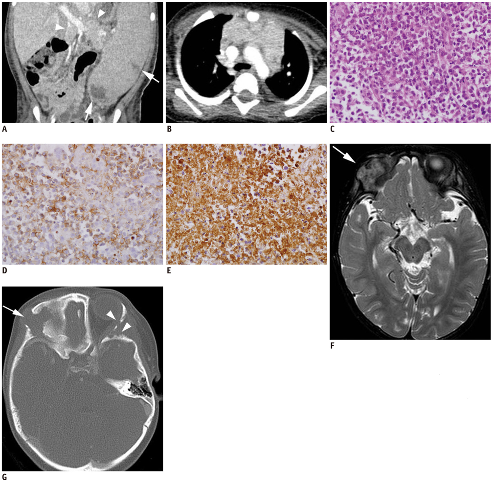
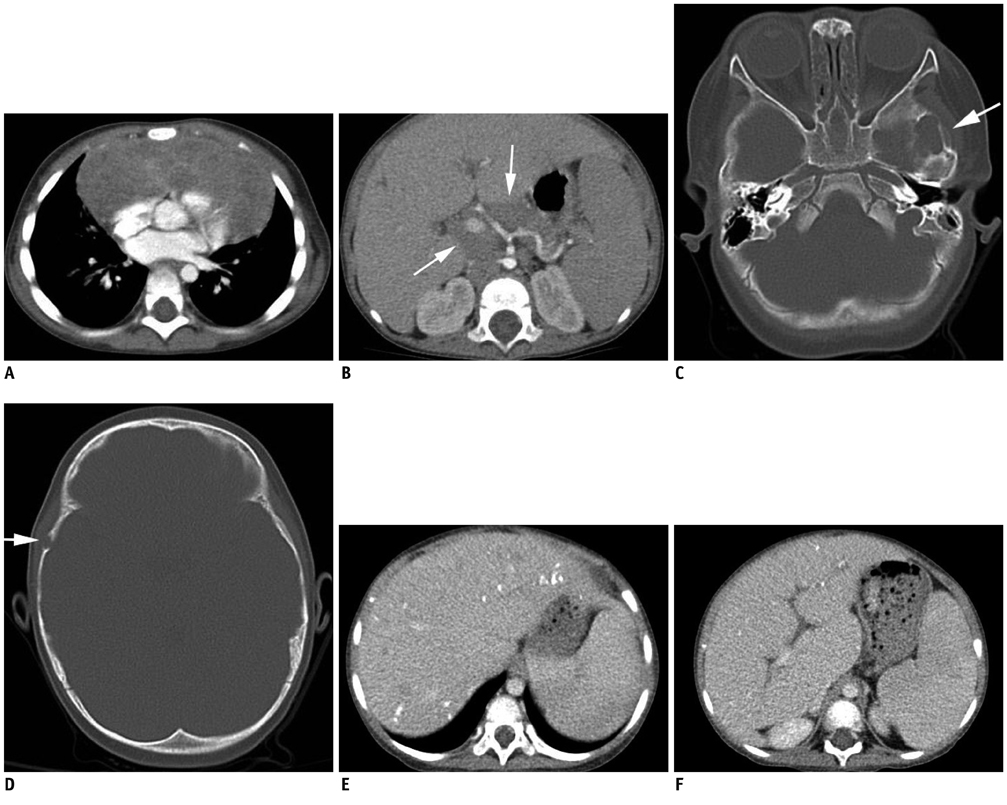
- Langerhans cell sarcoma (LCS) is a neoplastic proliferation of Langerhans cells with malignant cytological features and multi-organ involvement that typically has a poor prognosis. We experienced 2 cases of LCS in children less than 2 years of age and report them based primarily on CT and MR findings. Both children had findings of hepatosplenomegaly with low-attenuation nodular lesions, had multiple lymphadenopathy, and had shown recurrent lesions invading the skull during follow-up after chemotherapy.
Keyword
MeSH Terms
Figure
Cited by 1 articles
-
A Case of Langerhans Cell Sarcoma Presenting as Submandibular Gland Mass
Geonho Lee, Kunho Song, Ki Wan Park, Bon Seok Koo
Korean J Otorhinolaryngol-Head Neck Surg. 2019;62(9):520-523. doi: 10.3342/kjorl-hns.2018.00661.
Reference
-
1. Zhao G, Luo M, Wu ZY, Liu Q, Zhang B, Gao RL, et al. Langerhans cell sarcoma involving gallbladder and peritoneal lymph nodes: a case report. Int J Surg Pathol. 2009. 17:347–353.2. Lee JS, Ko GH, Kim HC, Jang IS, Jeon KN, Lee JH. Langerhans cell sarcoma arising from Langerhans cell histiocytosis: a case report. J Korean Med Sci. 2006. 21:577–580.3. Nakayama M, Takahashi K, Hori M, Okumura T, Saito M, Yamakawa M, et al. Langerhans cell sarcoma of the cervical lymph node: a case report and literature review. Auris Nasus Larynx. 2010. 37:750–753.4. Luo B, Pian B, Peng Z, Jintao D, Shixi L. Pharyngeal tonsil manifestation of Langerhans cells sarcoma: a case report and review of the literature. Int J Pediatr Otorhinolaryngol Extra. 2011. 6:156–158.5. Uchida K, Kobayashi S, Inukai T, Noriki S, Imamura Y, Nakajima H, et al. Langerhans cell sarcoma emanating from the upper arm skin: successful treatment by MAID regimen. J Orthop Sci. 2008. 13:89–93.6. Henter JI, Horne A, Aricó M, Egeler RM, Filipovich AH, Imashuku S, et al. HLH-2004: Diagnostic and therapeutic guidelines for hemophagocytic lymphohistiocytosis. Pediatr Blood Cancer. 2007. 48:124–131.7. Donadieu J, Chalard F, Jeziorski E. Medical management of langerhans cell histiocytosis from diagnosis to treatment. Expert Opin Pharmacother. 2012. 13:1309–1322.8. Caruso S, Miraglia R, Maruzzelli L, Luca A, Gridelli B. Biliary wall calcification in Langerhans cell histiocytosis: report of two cases. Pediatr Radiol. 2008. 38:791–794.9. Pyun HW, Kim ME, Kim JH. CT and MR findings of langerhans cell histiocytois involving the spleen: a case report. J Korean Radiol Soc. 2002. 46:171–174.10. Heller GD, Haller JO, Berdon WE, Sane S, Kleinman PK. Punctate thymic calcification in infants with untreated Langerhans' cell histiocytosis: report of four new cases. Pediatr Radiol. 1999. 29:813–815.11. Yi G, Yoon HK, Kim BK, Kim KA, Choo IW. CT findings of orbital langerhans cell histiocytosis. J Korean Radiol Soc. 2000. 42:841–846.
- Full Text Links
- Actions
-
Cited
- CITED
-
- Close
- Share
- Similar articles
-
- A Case of Langerhans Cell Sarcoma Presenting as Submandibular Gland Mass
- Clear Cell Sarcoma-Like Tumor of the Gastrointestinal Tract with Peritoneal Metastasis in a Young Adult: A Case Report with Literature Review
- Langerhans Cell Histiocytosis with Pancreatic Involvement: Imaging Findings Including Diffusion-Weighted Imaging
- A Case of Orbital Langerhans' cell histiocytosis
- Epithelioid Sarcoma: A Case Report


