A meningomyelocele with normal intracranial signs on ultrasound and false-negative amniotic fluid alpha-fetoprotein and acetylcholinesterase
- Affiliations
-
- 1Department of Obstetrics and Gynecology, Eulji University Medical Center, Eulji University College of Medicine, Daejeon, Korea. rho27kim@eulji.ac.kr
- 2Department of Neurosurgery, Eulji University Medical Center, Eulji University College of Medicine, Daejeon, Korea.
- KMID: 1704429
- DOI: http://doi.org/10.5468/ogs.2014.57.3.223
Abstract
- Neural tube defects are the major targets of prenatal diagnoses, along with Down syndrome. Prenatal diagnosis of spina bifida is possible at second trimester of gestation through alpha-fetoprotein and acetylcholinesterase biochemistry assays and ultrasound. In particular, the discovery of characteristic intracranial signs on ultrasound leads to a very high diagnosis rate. However, it is rare for spina bifida to present without intracranial signs while also showing normal values of maternal serum alpha-fetoprotein, amniotic fluid alpha-fetoprotein, and acetylcholinesterase. In our hospital, a fetus with spina bifida was delivered at 37+5 weeks' gestation by cesarean section, and was continually followed up over 2 years to date.
Keyword
MeSH Terms
-
Acetylcholinesterase*
alpha-Fetoproteins*
Amniotic Fluid*
Biochemistry
Cesarean Section
Diagnosis
Down Syndrome
Female
Fetus
Humans
Meningocele
Meningomyelocele*
Neural Tube Defects
Pregnancy
Pregnancy Trimester, Second
Prenatal Diagnosis
Reference Values
Spinal Dysraphism
Ultrasonography*
Acetylcholinesterase
alpha-Fetoproteins
Figure
-
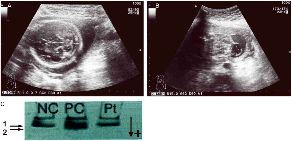
Fig. 1 (A,B) At 22+6 weeks' gestation, sonography shows normal range of cistern magna, no intracranial signs at axial view. Cystic mass is present in the lumbosacral area. (C) Native-polyacrylamide gel eletrophoresis analysis of amniotic fluid acetylcholinesterase was performed at 22+6 weeks' gestation. 1→ slow migrating thin band with specific acetylcholinesterase inhibitor; a negative control (lane NC). 2→ additional fast moving thick band of specific acetylcholinesterase; a positive control (lane PC). Lane Pt, patient shows a thin negative pattern.
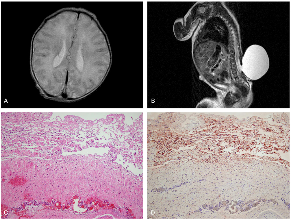
Fig. 2 (A) Immediate postnatal brain magnetic resonance imaging (MRI) shows normal findings. There is no lemon sign and no ventriculomegaly. (B) Spinal MRI. Sagittal T2-weighted MRI image of the lumbar meningocele. The level of defect was between L3 and L4. (C) The histopathologic examination revealed a meningocele (H&E, ×100). (D) Strong reactivity to epithelial membrane antigen (EMA) was seen in the epithelial lining and granules on the surface of the arachnoid and pia mater (EMA, ×100).
Reference
-
1. Nicolaides KH, Campbell S, Gabbe SG, Guidetti R. Ultrasound screening for spina bifida: cranial and cerebellar signs. Lancet. 1986; 2:72–74.2. Pierre-Kahn A, Sonigo P. Lumbosacral lipomas: in utero diagnosis and prognosis. Childs Nerv Syst. 2003; 19:551–554.3. Nievelstein RA, Hartwig NG, Vermeij-Keers C, Valk J. Embryonic development of the mammalian caudal neural tube. Teratology. 1993; 48:21–31.4. Boulet SL, Yang Q, Mai C, Kirby RS, Collins JS, Robbins JM, et al. Trends in the postfortification prevalence of spina bifida and anencephaly in the United States. Birth Defects Res A Clin Mol Teratol. 2008; 82:527–532.5. Kondo A, Kamihira O, Ozawa H. Neural tube defects: prevalence, etiology and prevention. Int J Urol. 2009; 16:49–57.6. Sebire NJ, Spencer K, Noble PL, Hughes K, Nicolaides KH. Maternal serum alpha-fetoprotein in fetal neural tube and abdominal wall defects at 10 to 14 weeks of gestation. Br J Obstet Gynaecol. 1997; 104:849–851.7. Loft AG, Hogdall E, Larsen SO, Norgaard-Pedersen B. A comparison of amniotic fluid alpha-fetoprotein and acetylcholinesterase in the prenatal diagnosis of open neural tube defects and anterior abdominal wall defects. Prenat Diagn. 1993; 13:93–109.8. Boogert A, Aarnoudse JG, De Bruijn HW, Gouw A. False-negative amniotic fluid acetylcholinesterase in a case of meningo-encephalocele. Prenat Diagn. 1989; 9:133–138.9. Harmon JP, Hiett AK, Palmer CG, Golichowski AM. Prenatal ultrasound detection of isolated neural tube defects: is cytogenetic evaluation warranted? Obstet Gynecol. 1995; 86(4 Pt 1):595–599.10. Watson WJ, Chescheir NC, Katz VL, Seeds JW. The role of ultrasound in evaluation of patients with elevated maternal serum alpha-fetoprotein: a review. Obstet Gynecol. 1991; 78:123–128.11. Tabor A, Vestergaard CH, Lidegaard O. Fetal loss rate after chorionic villus sampling and amniocentesis: an 11-year national registry study. Ultrasound Obstet Gynecol. 2009; 34:19–24.12. Cheschier N. ACOG Committee on Practice Bulletins-Obstetrics. ACOG practice bulletin: Neural tube defects. Number 44, July 2003 (Replaces committee opinion number 252, March 2001). Int J Gynaecol Obstet. 2003; 83:123–133.13. Bell JE, Gordon A, Maloney AF. The association of hydrocephalus and Arnold: Chiari malformation with spina bifida in the fetus. Neuropathol Appl Neurobiol. 1980; 6:29–39.14. Anteby EY, Yagel S. Route of delivery of fetuses with structural anomalies. Eur J Obstet Gynecol Reprod Biol. 2003; 106:5–9.
- Full Text Links
-
- Actions
-
Cited
- CITED
-
- Close
- Share
-
- Similar articles
-
- A case of false positive amniotic acetylcholinesterase in a normal pregnancy
- A Case of False Positive Amniotic Fluid Acetylcholinestserase in One Fetus of Twin Pregnancy conceived by Intracytoplasmic Sperm Injection and Zygote Intrafallopian Tube Transfer
- A Case of Spina Bifida Diagnosed by Prenatal Ultrasonography
- Amniotic Fluid Alpha-fetoprotein Levels in Midtrimester Pregnancies
- A Case of Positive Amniotic AFP, Acetylcholinesterase in a Normal Pregnancy after Undergoing Periodic Targeted Ultrasonographic Evaluation
