J Korean Soc Magn Reson Med.
2014 Mar;18(1):64-69. 10.13104/jksmrm.2014.18.1.64.
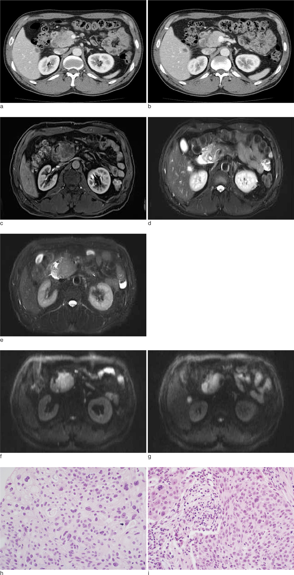
Pancreatic Adenosquamous Cell Carcinoma with Solitary Liver Metastasis Showing Different Imaging Features
- Affiliations
-
- 1Department of Radiology, Korea University Ansan Hospital, Korea University College of Medicine, Gyeonggi-do, Korea. sahcha@kumc.or.kr
- KMID: 1683782
- DOI: http://doi.org/10.13104/jksmrm.2014.18.1.64
Abstract
- Among exocrine pancreatic tumors, adenosquamous carcinoma is a rare, aggressive subtype with a poor prognosis and a high potential for metastases compared with its more conventional glandular counterpart, adenocarcinoma of the pancreas. We herein describe the imaging findings of pancreatic adenosquamous cell carcinoma with solitary liver metastasis showing different imaging features and also review the previous literature to recognize characteristic imaging features of pancreatic adenosquamous cell carcinoma.
Figure
Reference
-
1. Lampropoulos P, Filippous G, Skafida E, Vasilakaki T, Paschalidis N, Rizos S. Adenosquamous carcinoma of the pancreas, a rare tumor entity: a case report. Cases J. 2009; 2:9129.2. Trikudanathan G, Dasanu CA. Adenosquamous carcinoma of the pancreas: a distinct clinicopathologic entity. South Med J. 2010; 103:903–910.3. Ito K, Fujita N, Noda Y, et al. Pancreatic adenosquamous carcinoma, 9mm in size, diagnosed preoperatively by transpapillary biopsy: report of a case. Dig Endosc. 2003; 15:235–239.4. Nabae T, Yamaguchi K, Takahata S, et al. Adenosquamous carcinoma of the pancreas: report of two cases. Am J Gastroenterol. 1998; 93:1167–1170.5. Rahemtullah A, Misdraji J, Pitman MB. Adenosquamous carcinoma of the pancreas: cytologic features in 14 cases. Cancer. 2003; 99:372–378.6. Zhang B, Gao SL. Education of imaging. Hepatobiliary and pancreatic: adenosquamous carinoma of the pancreas. J Gastroenterol Hepatol. 2008; 23:820.7. Charbit A, Malaise EP, Tubiana M. Relation between the pathological nature and the growth rate of human tumors. Eur J Cancer. 1971; 7:307–315.8. Inoue T, Nagao S, Tajima H, et al. Adenosquamous pancreatic cancer producing parathyroid hormone-related protein. J Gastroenterol. 2004; 39:176–180.9. Komatsuda T, Ishida H, Konno K, et al. Adenosquamous carcinoma of the pancreas: report of two cases. Abdom Imaging. 2000; 25:420–423.10. Na YJ, Shim KN, Cho MS, et al. Primary adenosquamous cell carinoma of the pancreas: a case report with a review of the Korean literature. Korean J Intern Med. 2011; 26:348–351.
- Full Text Links
- Actions
-
Cited
- CITED
-
- Close
- Share
- Similar articles
-
- A Case of Cutaneous Metastatic Adenosquamous Carcinoma of the Pancreas
- A case of primary adenosquamous carcinoma of the liver
- Metastatic Cutaneous Adenosquamous Carcinoma in a Patient Undergoing Chemotherapy for Lung Adenocarcinoma
- An adenosquamous carcinoma of the liver that developed metachronously in a patient with a colon adenocarcinoma
- Adenosquamous Carcinoma of the Pancreas


