SAPHO Syndrome in a Patient with Breast Cancer Mimicking Bone Metastasis: A Case Report
- Affiliations
-
- 1Department of Radiology, Center for Breast Cancer, National Cancer Center of Korea, Korea. kokr@ncc.re.kr
- 2Department of Radiology, Center for Cancer Prevention and Early Detection, National Cancer Center of Korea, Korea.
- 3Department of Surgery, Center for Breast Cancer, National Cancer Center of Korea, Korea.
- 4Department of Pathology, Center for Breast Cancer, National Cancer Center of Korea, Korea.
- 5Department of Radiology, Center for Liver Cancer, National Cancer Center of Korea, Korea.
- KMID: 1683781
- DOI: http://doi.org/10.13104/jksmrm.2014.18.1.59
Abstract
- A 66-year-old woman was transferred to our hospital due to her right breast cancer. Preoperative breast MRI shows 1.9 cm malignancy on her right breast (cT1N0M0) and incidentally found osteosclerotic change of left coststernoclavicular region. Bone scintigraphy showed hot uptake and the possibility of bone metastasis was not excluded. However, because the bone metastasis is not common in early stage cancer and the costosternoclavicular region is not common site, other possibility should be considered. SAPHO syndrome can be diagnosed even in the absence of dermatosis when there is an axial or appendicular osteitis and hyperostosis, especially in costosternoclavicular region. Though breast imaging specialists are not accustomed to this disease entity, awareness and diagnosis of the SAPHO syndrome can help differentiate bone metastasis.
MeSH Terms
Figure
-
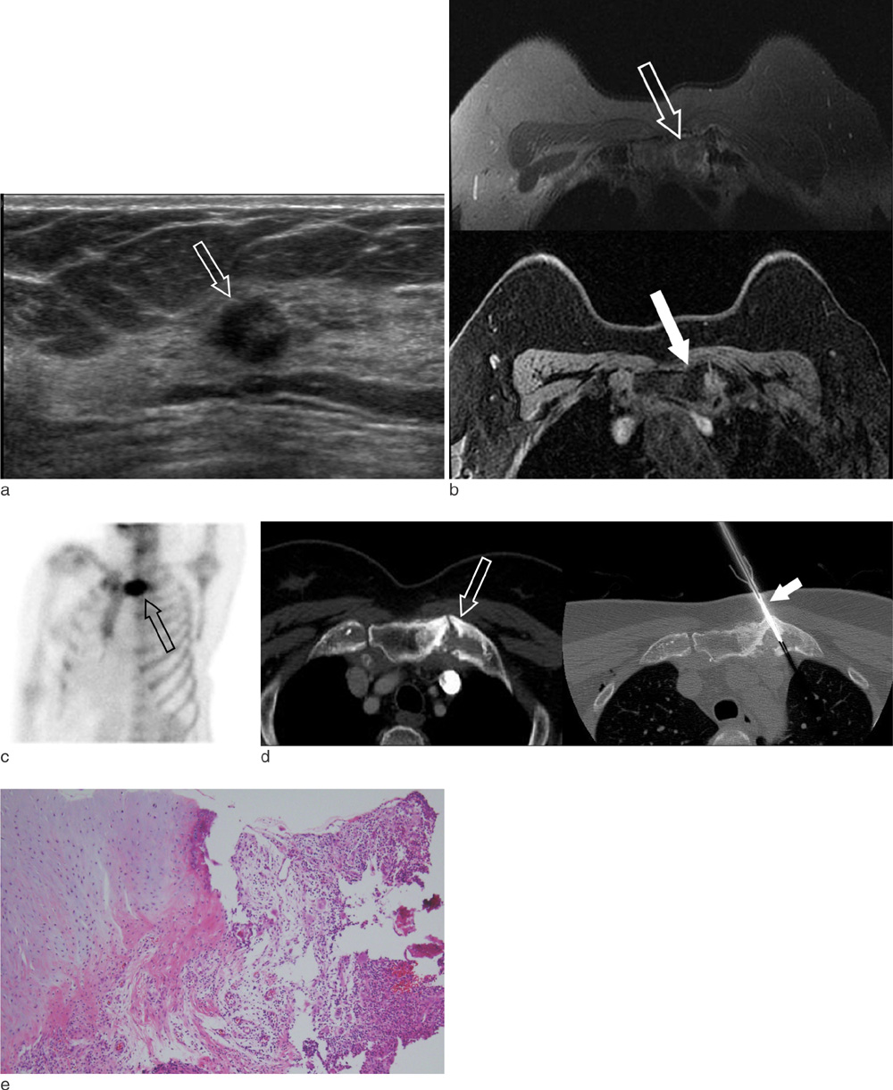
Fig. 1 Imaging findings in a 66-year-old woman with her right breast cancer. a. On breast ultrasound, about 1.3 cm-sized irregular mass is seen (hollow arrow) at her right breast which was confirmed as an invasive ductal carcinoma via gun biopsy. b. Breast MRI shows sclerotic bone changes of her left sternoclavicular joint (hollow arrows). On T2 weighted image, mild high signal intensities on the marrows of her left clavicle and manubrium (hollow arrow) are accompanied (top image) and on T1 weighted image, sclerotic cortical change is seen (white arrow, bottom image). c. Bone scintigraphy shows hot uptake at left costosternoclavicular region (hollow arrow). d. The osteosclerotic changes of left sternoclavicular region are seen on CT (right side, hollow arrow). To obtain the pathology, a bone biopsy needle (left side, white arrow) was advanced under CT guidance and chronic active inflammation without causative organism was confirmed. e. The chondro-osseous tissue is infiltrated by many inflammatory cells mainly composed of neutrophils and lymphocytes (× 100, H & E stain).
Reference
-
1. Hamaoka T, Madewell JE, Podoloff DA, Hortobagyi GN, Ueno NT. Bone imaging in metastatic breast cancer. J Clin Oncol. 2004; 22:2942–2953.2. Coleman RE, Rubens RD. The clinical course of bone metastases from breast cancer. Br J Cancer. 1987; 55:61–66.3. Hortobagyi GN. Bone metastases in breast cancer patients. Semin Oncol. 1991; 18:11–15.4. Boutin RD, Resnick D. The sapho syndrome: an evolving concept for unifying several idiopathic disorders of bone and skin. AJR Am J Roentgenol. 1998; 170:585–591.5. Rubens RD. Bone metastases--the clinical problem. Eur J Cancer. 1998; 34:210–213.6. Reith JD, Bauer TW, Schils JP. Osseous manifestations of sapho (synovitis, acne, pustulosis, hyperostosis, osteitis) syndrome. Am J Surg Pathol. 1996; 20:1368–1377.7. Assmann G, Simon P. The sapho syndrome--are microbes involved? Best Pract Res Clin Rheumatol. 2011; 25:423–434.8. Chamot AM, Vion B, Gerster JC. Acute pseudoseptic arthritis and palmoplantar pustulosis. Clin Rheumatol. 1986; 5:118–123.9. Sonozaki H, Mitsui H, Miyanaga Y, et al. Clinical features of 53 cases with pustulotic arthro-osteitis. Ann Rheum Dis. 1981; 40:547–553.10. Hellmann DB. Spondyloarthropathy with hidradenitis suppurativa. JAMA. 1992; 267:2363–2365.
- Full Text Links
-
- Actions
-
Cited
- CITED
-
- Close
- Share
-
- Similar articles
-
- A Case of SAPHO Syndrome Associated with Lytic Bone Lesions Resembling Metastases
- Fibrous Dysplasia Mimicking Bone Metastasis on Both Bone Scintigraphy and 18F-FDG PET-CT: Diagnostic Dilemma in a Patient with Breast Cancer
- SAPHO (Synovitis, Acne, Pustulosis, Hyperostosis, Osteitis) Syndrome occurred on the Clavicle: A Case Report
- Breast Cancer in Cowden Syndrome: A Case Report
- A case of stomach metastasis from breast cancer
