Perforator Flaps in Head and Neck Reconstruction
- Affiliations
-
- 1Department of Plastic and Reconstructive Surgery, School of Medicine, Hanyang University, Seoul, Korea. jtkim@hanyang.ac.kr
- KMID: 1505103
- DOI: http://doi.org/10.7599/hmr.2009.29.3.265
Abstract
- Basic requirements of head and neck reconstructions are thin resurfacing, a long vascular pedicle, 3-dimensional and well customized reconstruction with a team approach. Ideal reconstruction methods were thought to be free tissue transfer including radial forearm flap, latissmus dorsi or rectus abdominis myocutaneous flap. But recently, there has been concerns about sacrifice of donor structures in these conventional flaps. For minimal sacrifice of donor structures, there has been much evolution in flap concepts, which lead to the introduction of perforator flaps. They are popular in every region for reconstruction. Anterolatral thigh, latissmus dorsi or deep inferior epigastric artery perforator flaps are commonly used. Perforator flaps could also be applied to head and neck reconstructions, because they could be used for the controlled resurfacing of scalp, cheek, neck, oropharynx, and for customized 3-dimensional reconstructions, including diverse components according to each perforator, which may result in more comfortable handling and less restricted access to the defect. The perforator flaps also have long vascular pedicles compared to conventional myocutaneous flaps, which can lead to less restriction in choosing recipient vessels. Perforator flaps have known to have many advantages as described and they give one more option in head and neck reconstruction.
MeSH Terms
Figure
-
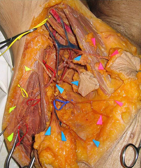
Fig. 1 Cadaver dissection shows three groups of perforator in axilla area. First rows(yellow) indicate musculotaneous perforator group, mid rows(blue) indicate septocutaneous perforator group and the other anterior rows(pink) indicate direct cutaneous group.
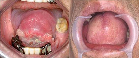
Fig. 2 (Left) preoperative view of tongue cancer. (Right) postoperative view of tongue resurfacing using perforator flap.
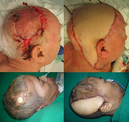
Fig. 3 (Above, Left) Recurrent squamous cell carcinoma in scalp. (Above, Right) 28×16 cm thoracodorsal artery perforator flap was used for resurfacing. (Below, Left) Preoperative view of skin defect and MRSA infection after cranioplasty. (Below, Right) A thoracodorsal artery perforator flap was used for the resurfacing of scalp defects.
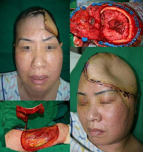
Fig. 4 (Above, Left) Recurrent MRSA infection of artificial dura and cerebritis (Above, Right) Intraoperative view of cranium (Below, Left) Vastus Lateralis perforator flap which is known as anterolateral thigh perforator flap was elevated. (Below, Right) postoperative view of scalp resurfacing.

Fig. 5 (Above, Left) Old male patient had suffered pharyngeal cancer with fistula formation and huge chyle leakage. (Above, Right) A septocutaneous perforator flap for pharyngeal lining and a musculocutaneous perforator skin flap for outer lining were elevated in anterolateral thigh. (Below, Left) Flap insetting for customized reconstruction. (Below, Right) Postoperative view shows successful reconstruction of pharyngeal lining and outer resurfacing.

Fig. 6 (Above, Left) total pharyngolaryngectomy state. (Above, Right) tubed latissimus dorsi perforator flap was elevated for cervical esophageal reconstruction. (Below, Left) Flap insetting for neoesophagus. (Below, Right) Postoperative endoscopic view shows successful reconstruction of esophagus.
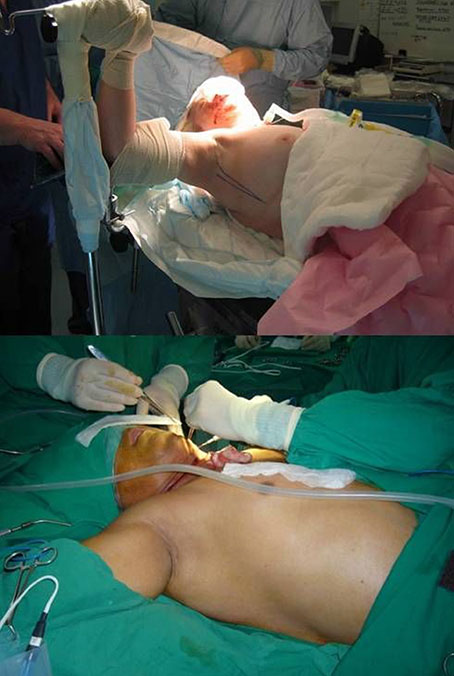
Fig. 7 (Above and Below) Generally latissimus dorsi or thracodorsal artery perforator flap is located more anterior to conventional muscle flap therefore you can harvest these flaps on supine position for two team approach.
Cited by 1 articles
-
Perspectives on reconstructive microsurgery in Korea
Byung-Joon Jeon, Goo-Hyun Mun
J Korean Med Assoc. 2011;54(6):604-616. doi: 10.5124/jkma.2011.54.6.604.
Reference
-
1. Lyons AJ. Perforator flaps in head and neck surgery. Int J Oral Maxillofac Surg. 2006; 35:199–207.
Article2. Koshima I, Yamamoto H, Hosoda M, Moriguchi T, Orita Y, Nagayama H. Free combined composite flaps using the lateral circumflex femoral system for repair of massive defects of the head and neck regions: an introduction to the chimeric flap principle. Plast Reconstr Surg. 1993; 92:411–420.
Article3. Koshima I, Fukuda H, Yamamoto H, Moriguchi T, Soeda S, Ohta S. Free anterolateral thigh flaps for reconstruction of head and neck defects. Plast Reconstr Surg. 1993; 92:421–428.
Article4. Kimata Y, Uchiyama K, Ebihara S, Yoshizumi T, Asai M, Saikawa M, Hayashi R, Jitsuiki Y, Majima K, Ohyama W, Haneda T, Nakatsuka T, Harii K. Versatility of the free anterolateral thigh flap for reconstruction of head and neck defects. Arch Otolaryngol Head Neck Surg. 1997; 123:1325–1331.
Article5. Anthony JP, Singer MI, Mathes SJ. Pharyngoesophageal reconstruction using the tubed free radial forearm flap. Clin Plast Surg. 1994; 21:137–147.
Article6. Scharpf J, Esclamado RM. Reconstruction with radial forearm flaps after ablative surgery for hypopharyngeal cancer. Head Neck. 2003; 25:261–266.
Article7. Sardesai MG, Fung K, Yoo JH, Bakker H. Donor-site morbidity following radial forearm free tissue transfer in head and neck surgery. J Otolaryngol Head Neck Surg. 2008; 37:411–416.8. O'Connell DA, Rieger J, Harris JR, Dziegielewski P, Zalmanowitz J, Sytsanko A, Li S, Wolfaardt J, Hart RD, Seikaly H. Swallowing function in patients with base of tongue cancers treated with primary surgery and reconstructed with a modified radial forearm free flap. Arch Otolaryngol Head Neck Surg. 2008; 134:857–864.9. Agrawal A, Husein OF, Schuller DE. Esophageal reconstruction with larynx preservation using forearm-free flap. Laryngoscope. 2008; 118:1750–1752.
Article10. Andrades P, Pehler SF, Baranano CF, Magnuson JS, Carroll WR, Rosenthal EL. Fistula analysis after radial forearm free flap reconstruction of hypopharyngeal defects. Laryngoscope. 2008; 118:1157–1163.
Article11. Piantanida R, Roselli R, Pellini R, Ferrario F, Boschini P, Spriano G. Reconstruction of major orbital-maxillary defects with free latissimus dorsi myocutaneous flap. Facial Plast Surg. 1999; 15:297–302.
Article12. Har-El G, Bhaya M, Sundaram K. Latissimus dorsi myocutaneous flap for secondary head and neck reconstruction. Am J Otolaryngol. 1999; 20:287–293.
Article13. Yamamoto Y, Nohira K, Yamashita T, Shintomi Y, Hosokawa M, Furkawa H, Sugihara T, Ohura T. Combined V figure-shaped scapular osteocutaneous and latissimus dorsi myocutaneous flap for composite mandibular reconstruction. Head Neck. 1995; 17:219–225.
Article14. Schliephake H, Schmelzeisen R, Neukam FW. The free revascularized rectus abdominis myocutaneous flap for the repair of tumour related defects in the head and neck area. Br J Oral Maxillofac Surg. 1996; 34:18–22.
Article15. Kyutoku S, Tsuji H, Inoue T, Kawakami K, Han F, Ogawa Y. Experience with the rectus abdominis myocutaneous flap with vascularized hard tissue for immediate orbitofacial reconstruction. Plast Reconstr Surg. 1999; 103:395–402.
Article16. Wanamaker JR, Burkey BB. Overview of the rectus abdominis myocutaneous flap in head and neck reconstruction. Facial Plast Surg. 1996; 12:45–50.
Article17. Markowitz BL, Satterberg T, Calcaterra T, Orringer J, Cohen S, Burstein F, Shaw W. The deep inferior epigastric rectus abdominis muscle and myocutaneous free tissue transfer: further applications for head and neck reconstruction. Ann Plast Surg. 1991; 27:577–582.
Article18. Koshima I, Soeda S. Inferior epigastric artery skin flaps without rectus abdominis muscle. Br J Plast Surg. 1989; 42:645–648.
Article19. Saint-Cyr M, Schaverien M, Wong C, Nagarkar P, Arbique G, Brown S, Rohrich RJ. The extended anterolateral thigh flap: anatomical basis and clinical experience. Plast Reconstr Surg. 2009; 123:1245–1255.
Article20. Yazar S, Gideroglu K, Kilic B, Gokkaya A. Use of composite anterolateral thigh flap as double-vascularised layers for reconstruction of complex hand dorsum defect. J Plast Reconstr Aesthet Surg. 2008; 61:1549–1550.
Article21. Kimura N, Saito M, Sumiya Y, Itoh N. Reconstruction of hand skin defects by microdissected mini anterolataral thigh perforator flaps. J Plast Reconstr Aesthet Surg. 2008; 61:1073–1077.
Article22. Kuo YR, Jeng SF, Wei FC, Su CY, Chien CY. Functional reconstruction of complex lip and cheek defect with free composite anterolateral thigh flap and vascularized fascia. Head Neck. 2008; 30:1001–1006.
Article23. Wei FC, Celik N, Jeng SF. Application of "simplified nomenclature for compound flaps" to the anterolateral thigh flap. Plast Reconstr Surg. 2005; 115:1051–1055.
Article24. Kim JT. Two options for perforator flaps in the flank donor site: latissimus dorsi and thoracodorsal perforator flaps. Plast Reconstr Surg. 2005; 115:755–763.
Article25. Kim JT. Latissimus dorsi perforator flap. Clin Plast Surg. 2003; 30:403–431.
Article26. Kim JT, Kim SK. Hand resurfacing with the superthin latissimus dorsi perforator-based free flap. Plast Reconstr Surg. 2003; 111:366–370.
Article27. Blondeel PN, Boeckx WD. Refinements in free flap breast reconstruction: the free bilateral deep inferior epigastric perforator flap anastomosed to the internal mammary artery. Br J Plast Surg. 1994; 47:495–501.
Article28. Hamdi M, Blondeel P, Van Landuyt K, Tondu T, Monstrey S. Bilateral autogenous breast reconstruction using perforator free flaps: a single center's experience. Plast Reconstr Surg. 2004; 114:83–89.
Article29. Koshima I, Inagawa K, Urushibara K, Moriguchi T. Paraumbilical perforator flap without deep inferior epigastric vessels. Plast Reconstr Surg. 1998; 102:1052–1057.
Article30. Koshima I, Inagawa K, Urushibara K, Ohtsuki M, Moriguchi T. Deep inferior epigastric perforator dermal-fat or adiposal flap for correction of craniofacial contour deformities. Plast Reconstr Surg. 2000; 106:10–15.
Article31. Koshima I, Nanba Y, Tsutsui T, Takahashi Y, Watanabe A, Ishii R. Free perforator flap for the treatment of defects after resection of huge arteriovenous malformations in the head and neck regions. Ann Plast Surg. 2003; 51:194–199.
Article32. Kim JT. New nomenclature concept of perforator flap. Br J Plast Surg. 2005; 58:431–440.
Article33. Imai R, Matsumura H, Tanaka K, Uchida R, Watanabe K. Comparison of Doppler sonography and multidetector-row computed tomography in the imaging findings of the deep inferior epigastric perforator artery. Ann Plast Surg. 2008; 61:94–98.
Article34. Mun GH, Kim HJ, Cha MK, Kim WY. Impact of perforator mapping using multidetector-row computed tomographic angiography on free thoracodorsal artery perforator flap transfer. Plast Reconstr Surg. 2008; 122:1079–1088.
Article35. Masia J, Clavero JA, Larrañaga JR, Alomar X, Pons G, Serret P. Multidetector-row computed tomography in the planning of abdominal perforator flaps. J Plast Reconstr Aesthet Surg. 2006; 59:594–599.
Article36. Mardini S, Tsai FC, Wei FC. The thigh as a model for free style flaps. Clin Plast Surg. 2003; 30:473–480.37. Coleman JJ 3rd, Tan KC, Searles JM, Hester TR, Nahai F. Jejunal free autograft: analysis of complications and their resolution. Plast Reconstr Surg. 1989; 84:589–595.38. Schusterman MA, Shestak K, de Vries EJ, Swartz W, Jones N, Johnson J, Myers E, Reilly J Jr. Reconstruction of the cervical esophagus: free jejunal transfer versus gastric pull-up. Plast Reconstr Surg. 1990; 85:16–21.39. Reece GP, Schusterman MA, Miller MJ, Kroll SS, Robb GL, Baldwin BJ, Luethcke DR. Morbidity and functional outcome of free jejunal transfer reconstruction for circumferential defects of the pharynx and cervical esophagus. Plast Reconstr Surg. 1995; 96:1307–1316.
Article40. Cordeiro PG, Shah K, Santamaria E, Gollub MJ, Singh B, Shah JP. Barium swallows after free jejunal transfer: should they be performed routinely? Plast Reconstr Surg. 1999; 103:1167–1175.
Article41. Yu P, Robb GL. Pharyngoesophageal reconstruction with the anterolateral thigh flap: a clinical and functional outcomes study. Plast Reconstr Surg. 2005; 116:1845–1855.
Article
- Full Text Links
-
- Actions
-
Cited
- CITED
-
- Close
- Share
-
- Similar articles
-
- Contribution of Perforator Flaps in the Flap Selection for Head and Neck Reconstruction
- Various Utility of Perforator Flaps in Head and Neck Reconstruction
- Perforating patterns of cutaneous perforator vessels in anterolateral thigh flaps for head and neck reconstruction and clinical outcomes
- Concept of perforator flap and reconstruction using microsurgery
- Perforator Flap versus Conventional Flap
