Korean J Ophthalmol.
2013 Apr;27(2):141-144. 10.3341/kjo.2013.27.2.141.
Increased Intraocular Pressure after Extensive Conjunctival Removal: A Case Report
- Affiliations
-
- 1Department of Ophthalmology, Asan Medical Center, University of Ulsan College of Medicine, Seoul, Korea. sungeye@gmail.com
- KMID: 1503809
- DOI: http://doi.org/10.3341/kjo.2013.27.2.141
Abstract
- A 50-year-old woman, who had undergone extensive removal of conjunctiva on the right eye for cosmetic purposes at a local clinic 8 months prior to presentation, was referred for uncontrolled intraocular pressure (IOP) elevation (up to 38 mmHg) despite maximal medical treatment. The superior and inferior conjunctival and episcleral vessels were severely engorged and the nasal and temporal bulbar conjunctival areas were covered with an avascular epithelium. Gonioscopic examination revealed an open angle with Schlemm's canal filled with blood to 360 degrees in the right eye. Brain and orbital magnetic resonance imaging and angiography results were normal. With the maximum tolerable anti-glaucoma medications, the IOP gradually decreased to 25 mmHg over 4 months of treatment. Extensive removal of conjunctiva and Tenon's capsule, leaving bare sclera, may lead to an elevation of the episcleral venous pressure because intrascleral and episcleral veins may no longer drain properly due to a lack of connection to Tenon's capsule and the conjunctival vasculature. This rare case suggests one possible mechanism of secondary glaucoma following ocular surgery.
MeSH Terms
Figure
-
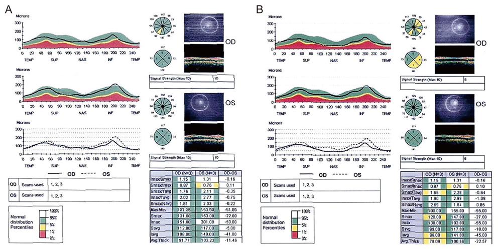
Fig. 1 Optical coherence tomography retinal nerve fiber layer (RNFL) scans obtained (A) preoperatively, and (B) 4 months after surgery. Average RNFL thickness in the right eye fell from 91.77 microns to 78.09 microns.

Fig. 2 (A,B) Anterior segment examination of the right eye, demonstrating episcleral and conjunctival engorgement in the superior and inferior areas. (C) The temporal bulbar conjunctiva lacked conjunctival vessels and was covered with a thin epithelium, with few engorged episcleral vessels. (D) Gonioscopic examination demonstrated an open angle with blood in Schlemm's canal.
Reference
-
1. Rhee DJ, Gupta M, Moncavage MB, et al. Idiopathic elevated episcleral venous pressure and open-angle glaucoma. Br J Ophthalmol. 2009. 93:231–234.2. Ruiz-Ederra J, Verkman AS. Mouse model of sustained elevation in intraocular pressure produced by episcleral vein occlusion. Exp Eye Res. 2006. 82:879–884.3. Shareef SR, Garcia-Valenzuela E, Salierno A, et al. Chronic ocular hypertension following episcleral venous occlusion in rats. Exp Eye Res. 1995. 61:379–382.4. Urcola JH, Hernandez M, Vecino E. Three experimental glaucoma models in rats: comparison of the effects of intraocular pressure elevation on retinal ganglion cell size and death. Exp Eye Res. 2006. 83:429–437.5. Bron AJ, Tripathi RC, Tripathi BJ, Wolff E. Wolff's anatomy of the eye and orbit. 1997. 8th ed. London: Chapman & Hall Medical;292–298.6. Mittag TW, Danias J, Pohorenec G, et al. Retinal damage after 3 to 4 months of elevated intraocular pressure in a rat glaucoma model. Invest Ophthalmol Vis Sci. 2000. 41:3451–3459.
- Full Text Links
-
- Actions
-
Cited
- CITED
-
- Close
- Share
-
- Similar articles
-
- The Effect of Ocusert for the Treatment of Glaucoma
- Surgical Removal of Sub-Tenon Triamcinolone Acetonide in Cases of Increased Intraocular Pressure after Sub-Tenon Injection
- A Case of Conjunctival Malignant Melanoma with Extensive Corneal Displacement
- A Case of Primary Conjunctival Giant Cell Tumor
- Encapsulated Bleb and Fibrous Ingrowth in the Ahmed Glaucoma Valve System and Bleb Revision with Mitomycin-C
