Clinical and Radiological Spectrum of Posterior Reversible Encephalopathy Syndrome
- Affiliations
-
- 1Department of Neurosurgery, Kangdong Sacred Heart Hospital, Hallym University College of Medicine, Seoul, Korea. nschbm@hallym.or.kr
- 2Department of Neurosurgery, Hallym University Sacred Heart Hospital, Hallym University College of Medicine, Anyang, Korea.
- KMID: 1491440
- DOI: http://doi.org/10.7461/jcen.2013.15.3.206
Abstract
OBJECTIVE
Symptoms of posterior reversible encephalopathy syndrome (PRES) include headache, altered mental status, visual disturbances, and seizures. Typical radiological features include edema of the parieto-occipital lobes. The purpose of this study is to review the clinical and radiological findings in patients diagnosed with PRES.
METHODS
All patients diagnosed with PRES between January 2006 and December 2012 were retrospectively included in this study. We reviewed demographic and clinical characteristics, and radiological findings.
RESULTS
We identified 16 patients with PRES. The most common clinical presentation was seizure (n = 12, 75%). Clinical recovery occurred in all patients within days (mean, 5.7 +/- 4.6 days). Comorbid conditions included hypertension (n = 4, 25%), cytotoxic medications (n = 3, 18.8%), sepsis (n = 4, 25%), malignancy (n = 4, 25%), subarachnoid hemorrhage (n = 1, 6.3%), autoimmune disorders (n = 1, 6.3%) and eclampsia (n = 1, 6.3%). The most commonly involved location was the parieto-occipital lobe (n = 13, 81.3%). Atypical radiological findings included significant basal ganglia involvement in 4 episodes; brainstem in 3, cerebellum in 2, and thalamus in 3. Eleven patients (68.8%) underwent diffusion-weighted imaging and apparent diffusion coefficient mapping. Of those, 9 patients (81.8%) had hypo- or isointensity on diffusion-weighted imaging. On the apparent diffusion coefficient map, 10 patients (90.9%) had hyperintensity, and the other had normal values.
CONCLUSION
We suggest that PRES may occur in patients with complex systemic conditions. The prognosis of PRES is usually benign. Physicians should be aware of certain atypical radiological findings to avoid a delayed diagnosis of PRES, as delayed diagnosis and treatment can result in permanent neurological sequlae.
MeSH Terms
Figure
-
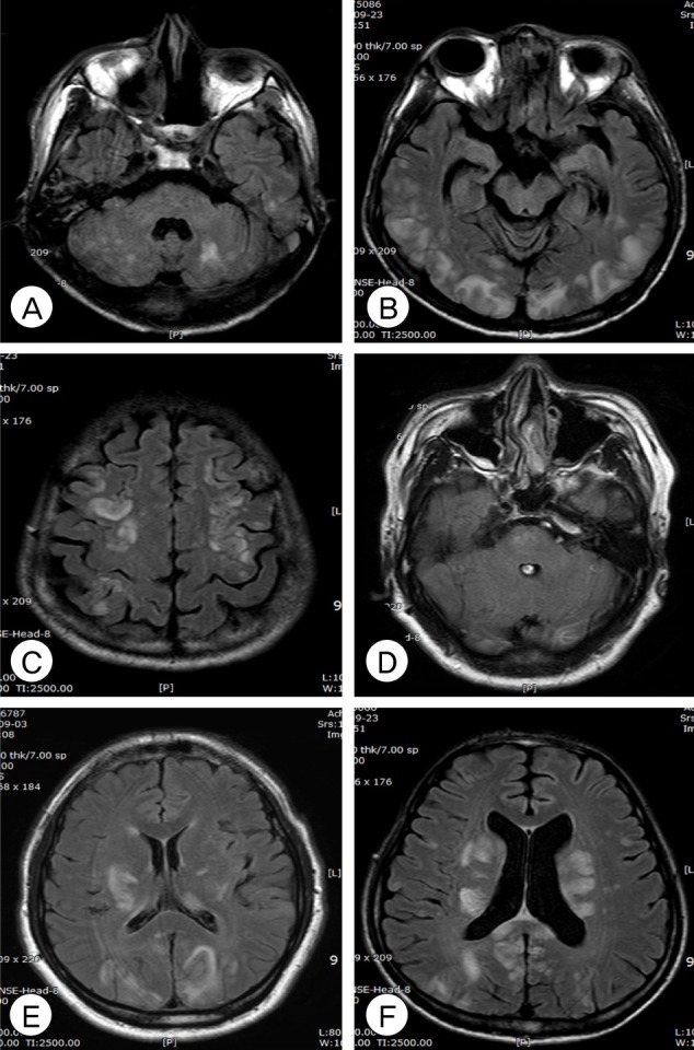
Fig. 1 Axial fluid-attenuated inversion recovery magnetic resonance imaging (MRI) of posterior reversible encephalopathy syndrome (PRES) shows the involvement of cerebellum (A), cortex (B), frontal lobe (C), brainstem with atypical pattern (D), and thalamus/basal ganglia (E, F).
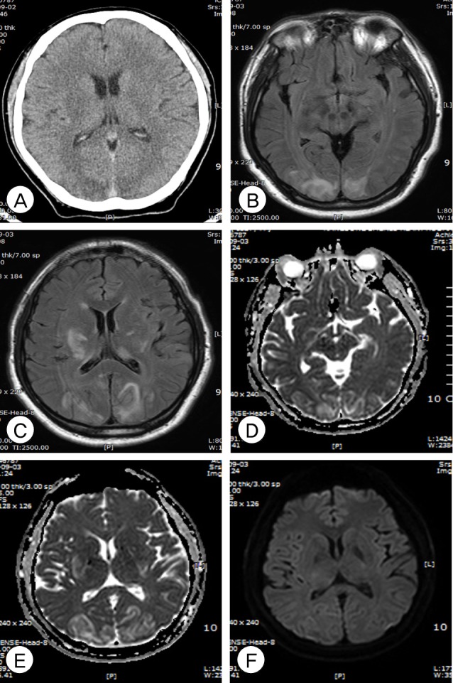
Fig. 2 A 43-year old female treated with cyclosporine-A (Patient 16). An axial computed tomography scan at onset shows low density at parieto-occipital lobes (A). Axial magnetic resonance imaging (MRI) shows a diffuse high-signal intensity lesion on fluid-attenuated inversion recovery MRI (B, C) and the apparent diffusion coefficient map (D, E) at the bilateral parieto-occipital lobes and basal ganglia. Diffusion weighted imaging shows lesions with iso and hypointensity (F) in the same distribution.
Cited by 1 articles
-
Evaluation and Treatment of the Acute Cerebral Infarction with Convexal Subarachnoid Hemorrhage
Min Hyung Lee, Sang Uk Kim, Dong Hoon Lee, Young Il Kim, Chul Bum Cho, Seung Ho Yang, Il Sup Kim, Jae Taek Hong, Jae Hoon Sung, Sang Won Lee
J Cerebrovasc Endovasc Neurosurg. 2016;18(3):271-275. doi: 10.7461/jcen.2016.18.3.271.
Reference
-
1. Ahn KJ, You WJ, Jeong SL, Lee JW, Kim BS, Lee JH, et al. Atypical manifestations of reversible posterior leukoencephalopathy syndrome: Findings on diffusion imaging and ADC mapping. Neuroradiology. 2004; 12. 46(12):978–983. PMID: 15536557.
Article2. Aird WC. The role of the endothelium in severe sepsis and multiple organ dysfunction syndrome. Blood. 2003; 5. 15. 101(10):3765–3777. PMID: 12543869.
Article3. Bartynski WS, Boardman JF. Catheter angiography, MR angiography, and MR perfusion in posterior reversible encephalopathy syndrome. AJNR Am J Neuroradiol. 2008; 3. 29(3):447–455. PMID: 18079186.
Article4. Bartynski WS, Boardman JF. Distinct imaging patterns and lesion distribution in posterior reversible encephalopathy syndrome. AJNR Am J Neuroradiol. 2007; 8. 28(7):1320–1327. PMID: 17698535.
Article5. Bartynski WS, Boardman JF, Zeigler ZR, Shadduck RK, Lister J. Posterior reversible encephalopathy syndrome in infection, sepsis, and shock. AJNR Am J Neuroradiol. 2006; Nov-Dec. 27(10):2179–2190. PMID: 17110690.6. Bartynski WS, Zeigler Z, Spearman MP, Lin L, Shadduck RK, Lister J. Etiology of cortical and white matter lesions in cyclosporin-A and FK-506 neurotoxicity. AJNR Am J Neuroradiol. 2001; Nov-Dec. 22(10):1901–1914. PMID: 11733324.7. Bhatt A, Farooq MU, Majid A, Kassab M. Chemotherapy-related posterior reversible leukoencephalopathy syndrome. Nat Clin Pract Neurol. 2009; 3. 5(3):163–169. PMID: 19262592.
Article8. Casey SO, McKinney A, Teksam M, Liu H, Truwit CL. CT perfusion imaging in the management of posterior reversible encephalopathy. Neuroradiology. 2004; 4. 46(4):272–276. PMID: 15045493.
Article9. Cooney MJ, Bradley WG, Symko SC, Patel ST, Groncy PK. Hypertensive encephalopathy: Complication in children treated for myeloproliferative disorders- Report of three cases. Radiology. 2000; 3. 214(3):711–716. PMID: 10715035.10. Dekker GA, Sibai BM. Etiology and pathogenesis of preeclampsia: Current concepts. Am J Obstet Gynecol. 1998; 11. 179(5):1359–1375. PMID: 9822529.
Article11. Domínguez-Fuentes B, Garcia-Gil D, Romero-Palacios A, Sanchez-Crespo JM, Garcia-Arjona R, Navarro-Navarro J. [Posterior reversible leukoencephalopathy in a patient with postpartum eclampsia]. Med Intensiva. 2008; 10. 32(7):361–363. Spanish. PMID: 18842228.12. Edvinsson L, Owman C, Sjoberg NO. Autonomic nerves, mast cells, and amine receptors in human brain vessels. A histochemical and pharmacological study. Brain Res. 1976; 10. 115(3):377–393. PMID: 184880.
Article13. Eichler FS, Wang P, Wityk RJ, Beauchamp NJ Jr, Barker PB. Diffuse metabolic abnormalities in reversible posterior leukoencephalopathy syndrome. AJNR Am J Neuroradiol. 2002; 5. 23(5):833–837. PMID: 12006287.14. Glusker P, Recht L, Lane B. Reversible posterior leukoencephalopathy syndrome and bevacizumab. N Engl J Med. 2006; 3. 354(9):980–982. discussion 980-2. PMID: 16510760.
Article15. Hefzy HM, Bartynski WS, Boardman JF, Lacomis D. Hemorrhage in posterior reversible encephalopathy syndrome: Imaging and clinical features. AJNR Am J Neuroradiol. 2009; 8. 30(7):1371–1379. PMID: 19386731.
Article16. Hinchey J, Chaves C, Appignani B, Breen J, Pao L, Wang A, et al. A reversible posterior leukoencephalopathy syndrome. N Engl J Med. 1996; 2. 334(8):494–500. PMID: 8559202.
Article17. Ito Y, Arahata Y, Goto Y, Hirayama M, Nagamutsu M, Yasuda T, et al. Cisplatin neurotoxicity presenting as reversible posterior leukoencephalopathy syndrome. AJNR Am J Neuroradiol. 1998; 3. 19(3):415–417. PMID: 9541291.18. Kinoshita T, Moritani T, Shrier DA, Hiwatashi A, Wang HZ, Numaguchi Y, et al. Diffusion-weighted MR imaging of posterior reversible leukoencephalopathy syndrome: A pictorial essay. Clin Imaging. 2003; Sep-Oct. 27(5):307–315. PMID: 12932680.19. Kitaguchi H, Tomimoto H, Miki Y, Yamamoto A, Terada K, Satoi H, et al. A brainstem variant of reversible posterior leukoencephalopathy syndrome. Neuroradiology. 2005; 9. 47(9):652–656. PMID: 15947925.
Article20. Kwon S, Koo J, Lee S. Clinical spectrum of reversible posterior leukoencephalopathy syndrome. Pediatr Neurol. 2001; 5. 24(5):361–364. PMID: 11516610.
Article21. Lassila M, Santisteban J, Finckenberg P, Salmenpera P, Riutta A, Moilanen E, et al. Vascular changes in cyclosporine A-induced hypertension and nephrotoxicity in spontaneously hypertensive rats on high-sodium diet. J Physiol Pharmacol. 2001; 3. 52(1):21–38. PMID: 11321510.22. Lee VH, Wijdicks EF, Manno EM, Rabinstein AA. Clinical spectrum of reversible posterior leukoencephalopathy syndrome. Arch Neurol. 2008; 2. 65(2):205–210. PMID: 18268188.
Article23. Lin JT, Wang SJ, Fuh JL, Hsiao LT, Lirng JF, Chen PM. Prolonged reversible vasospasm in cyclosporin A-induced encephalopathy. AJNR Am J Neuroradiol. 2003; 1. 24(1):102–104. PMID: 12533334.24. Min L, Zwerling J, Ocava LC, Chen IH, Putterman C. Reversible posterior leukoencephalopathy in connective tissue diseases. Semin Arthritis Rheum. 2006; 6. 35(6):388–395. PMID: 16765716.
Article25. Prasad N, Gulati S, Gupta RK, Kumar R, Sharma K, Sharma RK. Is reversible posterior leukoencephalopathy with severe hypertension completely reversible in all patients? Pediatr Nephrol. 2003; 11. 18(11):1161–1166. PMID: 14505162.
Article26. Primavera A, Audenino D, Mavilio N, Cocito L. Reversible posterior leucoencephalopathy syndrome in systemic lupus and vasculitis. Ann Rheum Dis. 2001; 5. 60(5):534–537. PMID: 11302882.
Article27. Pula JH, Eggenberger E. Posterior reversible encephalopathy syndrome. Curr Opin Ophthalmol. 2008; 11. 19(6):479–484. PMID: 18854692.
Article28. Schaefer PW, Gonzalez RG, Hunter G, Wang B, Koroshetz WJ, Schwamm LH. Diagnostic value of apparent diffusion coefficient hyperintensity in selected patients with acute neurologic deficits. J Neuroimaging. 2001; 10. 11(4):369–380. PMID: 11677876.
Article29. Schwartz RB, Bravo SM, Klufas RA, Hsu L, Barnes PD, Robson CD, et al. Cyclosporine neurotoxicity and its relationship to hypertensive encephalopathy: CT and MR findings in 16 cases. AJR Am J Roentgenol. 1995; 9. 165(3):627–631. PMID: 7645483.
Article30. Schwartz RB, Jones KM, Kalina P, Bajakian RL, Mantello MT, Garada B, et al. Hypertensive encephalopathy: Findings on CT, MR imaging, and SPECT imaging in 14 cases. AJR Am J Roentgenol. 1992; 8. 159(2):379–383. PMID: 1632361.
Article31. Servillo G, Bifulco F, De Robertis E, Piazza O, Striano P, Tortora F, et al. Posterior reversible encephalopathy syndrome in intensive care medicine. Intensive Care Med. 2007; 2. 33(2):230–236. PMID: 17119920.
Article32. Sharma A, Whitesell RT, Moran KJ. Imaging pattern of intracranial hemorrhage in the setting of posterior reversible encephalopathy syndrome. Neuroradiology. 2010; 10. 52(10):855–863. PMID: 19956935.
Article33. Trommer BL, Homer D, Mikhael MA. Cerebral vasospasm and eclampsia. Stroke. 1988; 3. 19(3):326–329. PMID: 3354016.
Article
- Full Text Links
-
- Actions
-
Cited
- CITED
-
- Close
- Share
-
- Similar articles
-
- Posterior Reversible Encephalopathy Syndrome
- Posterior Reversible Encephalopathy Syndrome in a Patient with Intoxication of Arisaema amurense
- A Case of Posterior Reversible Encephalopathy Syndrome in a Patient having Continuous Ambulatory Peritoneal Dialysis
- Malignant Posterior Reversible Encephalopathy Syndrome: A Case of Posterior Irreversible Encephalopathy Syndrome
- Posterior reversible encephalopathy syndrome and reversible cerebral vasoconstriction syndrome associated with acute exacerbation of chronic obstructive pulmonary disease
