Posterior reversible encephalopathy syndrome and reversible cerebral vasoconstriction syndrome associated with acute exacerbation of chronic obstructive pulmonary disease
- Affiliations
-
- 1Department of Neurology, Gyeongsang National University Changwon Hospital, Changwon, Korea
- 2Department of Neurology, Gyeongsang Institute of Health Science, College of Medicine, Gyeongsang National University, Jinju, Korea
- KMID: 2535721
- DOI: http://doi.org/10.14253/acn.2022.24.2.68
Abstract
- Posterior reversible encephalopathy syndrome (PRES) and reversible cerebral vasoconstriction syndrome (RCVS) are relatively uncommon neurological disorders. These two independent syndromes can be concurrent as a part of a continuum process; however, the specific mechanism is not well known. Although the relationship between RCVS and PRES is currently unclear, they could share a common pathophysiology. This case report aimed to determine the pathophysiology underlying the co-occurrence of PRES and RCVS in a patient with an acute exacerbation of chronic obstructive pulmonary disease.
Keyword
Figure
-
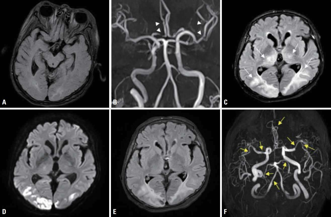
Fig. 1. Initial magnetic resonance imaging (MRI) was performed 2 days before admission. The axial section of a fluid-attenuated inversion recovery (FLAIR) image presents subtle hyperintensities in the bilateral occipital lobe (A). Magnetic resonance angiography (MRA) demonstrates multiple focal luminal narrowing (arrowheads) of the middle and anterior cerebral arteries (B). The second MRI was performed 5 days after admission. The axial FLAIR image presents newly developed high-signal-intensity lesions with parenchymal swelling in both parietooccipital lobes and both thalami (arrows) (C). Diffusion-weighted image presents diffusion restrictions in both medial frontal lobes and both parietooccipital lobes (mostly cortices) (D). The third MRI was performed 12 days after admission. The FLAIR image presents cortical and subcortical T2-weighted hyperintense lesions with parenchymal swelling in both medial frontal lobes and both parietooccipital lobes (E). MRA revealed multiple apparent stenoses in the following brain areas: both M1, M2, A3, both P2, BA, and V4 (arrows) (F).
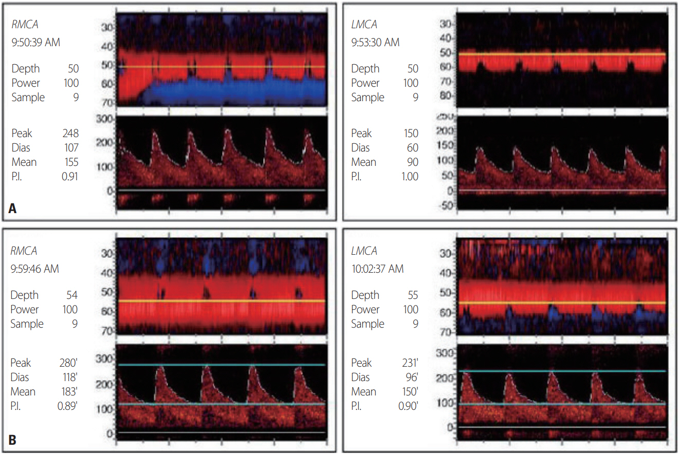
Fig. 2. Transcranial Doppler (TCD) ultrasonography was performed 16 days after admission. Increased flow velocities of 151-155 cm/sec were recorded in the right middle cerebral artery (RMCA), with a high Lindegaard ratio (3.88). Likewise, increased flow velocities of 90–95 cm/sec were recorded in the left middle cerebral artery (LMCA) (A). The second TCD ultrasonography investigations were performed 34 days after admission. Increased flow velocities of 183-191 cm/sec were recorded in the RMCA, with a high Lindegaard ratio (5.23). Likewise, increased flow velocities of 90-95 cm/sec were recorded in the RMCA, with a high Lindegaard ratio (3.51). Worsening of the pneumonia resulted in aggravation of carbon dioxide retention, which in turn worsened vasospasm (B). P.I., pulsatility index.
Reference
-
1. Valencia-Mendoza M, Ramírez-Rodríguez N, Vargas-Avila N, Peña-Ortiz A, Corzo-Villamizar M, Serna-Ramírez L, et al. Fatal reversible cerebral vasoconstriction syndrome: a systematic review of case series and case reports. J Clin Neurosci. 2019; 70:183–188.2. Hinchey J, Chaves C, Appignani B, Breen J, Pao L, Wang A, et al. A reversible posterior leukoencephalopathy syndrome. N Engl J Med. 1996; 334:494–500.3. Dardis C, Craciun R, Schell R. Posterior reversible encephalopathy syndrome in the setting of COPD: proposed pathogenesis. Med Hypotheses. 2013; 80:197–200.4. Khanal S, Acharya SP. Posterior reversible encephalopathy syndrome in association with exacerbation of chronic obstructive pulmonary disease: a case report. BMC Neurol. 2018; 18:1–3.5. Gupta H, Alrohimi A, Nathoo N, Nowacki T, Siddiqi ZA. Posterior reversible encephalopathy syndrome due to chronic obstructive pulmonary disease. Can J Neurol Sci. 2020; 47:569–571.6. Dietvorst S, Lambert J, Demeestere J, Lemmens R. Posterior reversible encephalopathy syndrome in a patient with chronic obstructive pulmonary disease. Acta Neurol Belg. 2020; 120:163–165.7. Sattar A, Manousakis G, Jensen MB. Systematic review of reversible cerebral vasoconstriction syndrome. Expert Rev Cardiovasc Ther. 2010; 8:1417–1421.
- Full Text Links
-
- Actions
-
Cited
- CITED
-
- Close
- Share
-
- Similar articles
-
- Reversible Cerebral Vasoconstriction Syndrome Combined with Posterior Encephalopathy Syndrome, and Transient Splenial Lesion after Delivery
- Reversible Cerebral Vasoconstriction Syndrome and Posterior Reversible Encephalopathy Syndrome Presenting with Deep Intracerebral Hemorrhage in Young Women
- Reversible Cerebral Vasoconstriction Syndrome in a Professional Rugby Player: A Case Report
- Posterior Reversible Encephalopathy Syndrome in a Patient with Intoxication of Arisaema amurense
- Reversible Cerebral Vasoconstriction Syndrome Induced by Blood Transfusion
