Korean J Radiol.
2013 Apr;14(2):233-239. 10.3348/kjr.2013.14.2.233.
Magnetic Resonance Evaluation of Mullerian Remnants in Mayer-Rokitansky-Kuster-Hauser Syndrome
- Affiliations
-
- 1Department of Radiology, Seoul National University Hospital, Seoul 110-744, Korea. radjycho@snu.ac.kr
- KMID: 1482782
- DOI: http://doi.org/10.3348/kjr.2013.14.2.233
Abstract
OBJECTIVE
To analyze magnetic resonance imaging (MRI) findings of Mullerian remnants in young females clinically suspected of Mayer-Rokitansky-Kuster-Hauser (MRKH) syndrome in a primary amenorrhea workup.
MATERIALS AND METHODS
Fifteen young females underwent multiplanar T2- and transverse T1-weighted MRI at either a 1.5T or 3.0T MR imager. Two gynecologic radiologists reached consensus decisions for the evaluation of Mullerian remnants, vagina, ovaries, and associated findings.
RESULTS
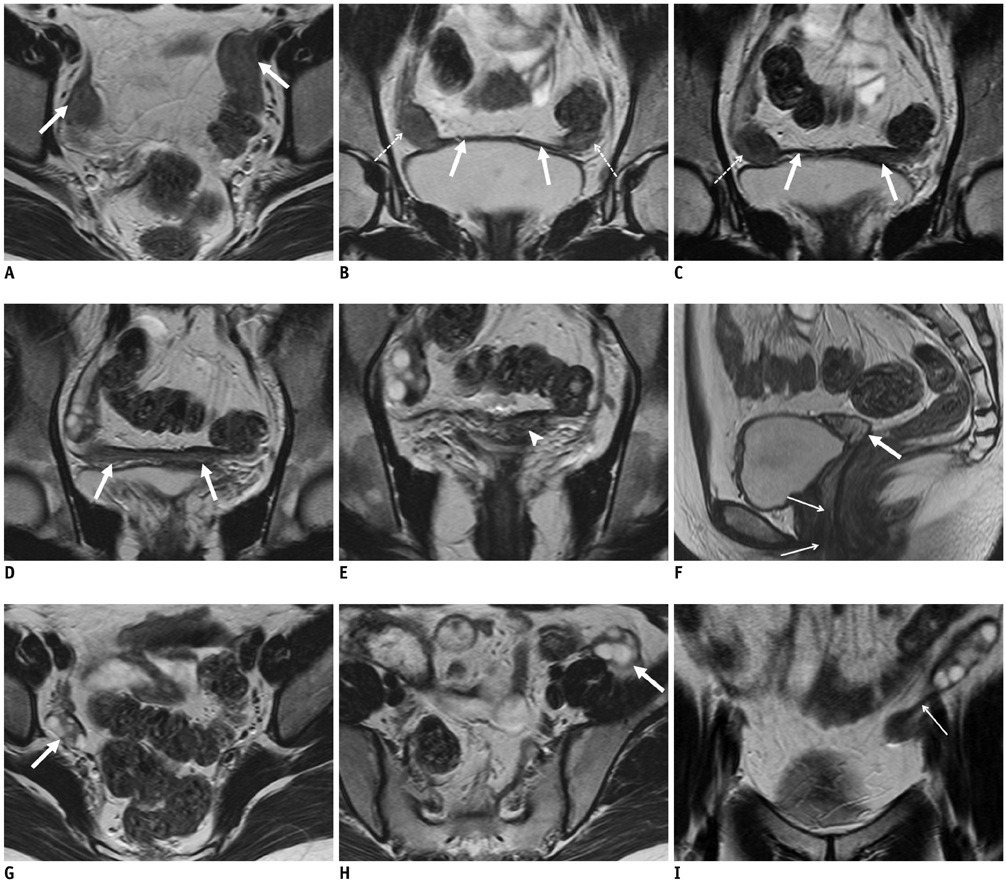
All cases had bilateral uterine buds in the pelvic cavity, with unilateral cavitation in two cases. The buds had an average long-axis diameter of 2.64 +/- 0.65 cm. In all cases, bilateral buds were connected with fibrous band-like structures. In 13 cases, the band-like structures converged at the midline or a paramedian triangular soft tissue lying above the bladder dome. The lower one-third of the vagina was identified in 14 cases. Fourteen cases showed bilateral normal ovaries near the uterine buds. One unilateral pelvic kidney, one unilateral renal agenesis, one mild scoliosis, and three lumbar sacralization cases were found as associated findings.
CONCLUSION
Typical Mullerian remnants in MRKH syndrome consist of bilateral uterine buds connected by the fibrous band-like structures, which converge at the midline triangular soft tissue lying above the bladder dome.
Keyword
MeSH Terms
-
Abnormalities, Multiple/*pathology
Adolescent
Adult
Female
Humans
Kidney/abnormalities/pathology
Magnetic Resonance Imaging/*methods
Middle Aged
Mullerian Ducts/abnormalities/pathology
Retrospective Studies
Somites/abnormalities/pathology
Spine/abnormalities/pathology
Uterus/abnormalities/pathology
Vagina/abnormalities/pathology
Figure
Reference
-
1. Giusti S, Fruzzetti E, Perini D, Fruzzetti F, Giusti P, Bartolozzi C. Diagnosis of a variant of Mayer-Rokitansky-Kuster-Hauser syndrome: useful MRI findings. Abdom Imaging. 2011. 36:753–755.2. Lamarca M, Navarro R, Ballesteros ME, García-Aguirre S, Conte MP, Duque JA. Leiomyomas in both uterine remnants in a woman with the Mayer-Rokitansky-Küster-Hauser syndrome. Fertil Steril. 2009. 91:931.e13–931.e15.3. Pompili G, Munari A, Franceschelli G, Flor N, Meroni R, Frontino G, et al. Magnetic resonance imaging in the preoperative assessment of Mayer-Rokitansky-Kuster-Hauser syndrome. Radiol Med. 2009. 114:811–826.4. Reinhold C, Hricak H, Forstner R, Ascher SM, Bret PM, Meyer WR, et al. Primary amenorrhea: evaluation with MR imaging. Radiology. 1997. 203:383–390.5. Zhou JH, Sun J, Yang CB, Xie ZW, Shao WQ, Jin HM. Long-term outcomes of transvestibular vaginoplasty with pelvic peritoneum in 182 patients with Rokitansky's syndrome. Fertil Steril. 2010. 94:2281–2285.6. Jurkiewicz B, Matuszewski L, Cisłak R, Rybak D. Rokitansky-Kustner-Hauser syndrome - a case report. Eur J Pediatr Surg. 2006. 16:135–137.7. Chandiramani M, Gardiner CA, Padfield CJ, Ikhena SE. Mayer - Rokitansky - Kuster - Hauser syndrome. J Obstet Gynaecol. 2006. 26:603–606.8. Deligeoroglou E, Kontoravdis A, Makrakis E, Christopoulos P, Kountouris A, Creatsas G. Development of leiomyomas on the uterine remnants of two women with Mayer-Rokitansky-Küster-Hauser syndrome. Fertil Steril. 2004. 81:1385–1387.9. Govindarajan M, Rajan RS, Kalyanpur A, Ravikumar . Magnetic resonance imaging diagnosis of Mayer-Rokitansky-Kuster-Hauser syndrome. J Hum Reprod Sci. 2008. 1:83–85.10. Jadoul P, Pirard C, Squifflet J, Smets M, Donnez J. Pelvic mass in a woman with Mayer-Rokitansky-Kuster-Hauser syndrome. Fertil Steril. 2004. 81:203–204.11. Lanowska M, Favero G, Schneider A, Köhler C. Laparoscopy for differential diagnosis of a pelvic mass in a patient with Mayer-Rokitanski-Küster-Hauser (MRKH) syndrome. Fertil Steril. 2009. 91:931.e17–931.e18.12. Papa G, Andreotti M, Giannubilo SR, Cesari R, Ceré I, Tranquilli AL. Case report and surgical solution for a voluminous uterine leiomyoma in a woman with complicated Mayer-Rokitansky-Küster-Hauser syndrome. Fertil Steril. 2008. 90:2014.e5–2014.e6.13. Sönmezer M, Atabekoglu C, Dökmeci F. Laparoscopic excision of symmetric uterine remnants in a patient with mayer-rokitansky-küster-hauser syndrome. J Am Assoc Gynecol Laparosc. 2003. 10:409–411.14. Chandler TM, Machan LS, Cooperberg PL, Harris AC, Chang SD. Mullerian duct anomalies: from diagnosis to intervention. Br J Radiol. 2009. 82:1034–1042.15. Reichman DE, Laufer MR. Mayer-Rokitansky-Küster-Hauser syndrome: fertility counseling and treatment. Fertil Steril. 2010. 94:1941–1943.
- Full Text Links
- Actions
-
Cited
- CITED
-
- Close
- Share
- Similar articles
-
- A Case of Uterine Leiomyomas in both Rudimentary Uterine Horns in a Woman with the Mayer-Rokitansky-Kuster-Hauser Syndrome
- A Case of Mayer-Rokitansky-Kuster-Hauser Syndrome Combined with Single Pelvic Kidney
- MR Findings of Mayer-Rokitansky-Kuster-Hauser Syndrome: Two Cases Report
- Uterine Adenomyosis Which Developed from Hypoplastic Uterus in Postmenopausal Woman with Mayer-Rokitansky-Kuster-Hauser Syndrome: A Case Report
- A Case of Mayer-Rokitansky-Kuster-Hauser (MRKH) Syndrome with Amenorrhea and Sexual Precosity


