Tuberc Respir Dis.
2008 Jan;64(1):28-32. 10.4046/trd.2008.64.1.28.
A Case of Pulmonary Arterial Thrombosis in a Patient with Tuberculous-destroyed Lung and Pulmonary Hypertension
- Affiliations
-
- 1Department of Internal Medicine, Pochon CHA University College of Medicine, Seongnam, Korea. jhcmd@hanmail.net
- 2Department of Radiology, Pochon CHA University College of Medicine, Seongnam, Korea.
- KMID: 1478138
- DOI: http://doi.org/10.4046/trd.2008.64.1.28
Abstract
- Pulmonary arterial thrombosis develops during hypercoagulable states, intra-arterial tumorous conditions, and congenital heart disease accompanied by pulmonary hypertension. Thrombosis in the main pulmonary arterial stump after pneumonectomy can also occur. Herein, we report a very rare case of pulmonary arterial thrombosis in a patient with pulmonary hypertension and a lung destroyed by tuberculosis. He presented with aggravated dyspnea without fever or purulent sputum. His chest computerized tomography scan showed left main pulmonary arterial thrombosis as a convex shape, with the ipsilateral distal arteries and arterioles showing parenchymal destruction. After excluding pulmonary thromboembolism and hypercoagulable disorders, we diagnosed pulmonary arterial thrombosis and treated him with an anticoagulant.
MeSH Terms
Figure
-
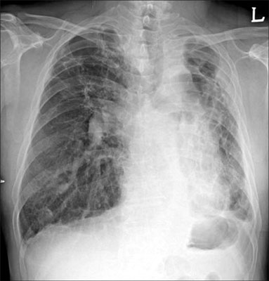
Figure 1 Chest X-ray on the first day at the hospital shows increased opacity in left lung with traction bronchiectasis and multiple calcified nodules.
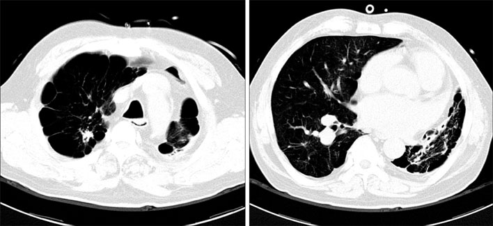
Figure 2 Lung setting of chest CT shows decreased lung volume, bullous emphysema, traction bronchiectasis, architectural distortion and multiple small calcifications in right upper lobe, superior segment of right lower lobe and left lung.

Figure 3 Contrast enhanced chest CT on the first day shows focal filling defect with convex-shape thrombus in left main pulmonary artery.
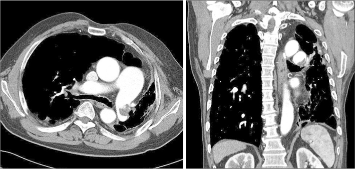
Figure 4 Contrast enhanced chest CT after anticoagulation for 2 weeks shows decreased size of thrombus in left main pulmonary artery.
Reference
-
1. Engelke C, Schaefer-Prokop C, Schirg E, Freihorst J, Grubnic S, Prokop M. High resolution CT and CT angiography of peripheral pulmonary vascular disorders. Radiographics. 2002. 22:739–764.2. Engelke C, Riedel M, Rummeny EJ, Marten K. Pulmonary haemangiosarcoma with main pulmonary artery thrombosis imitating subacute pulmonary embolism with infarction. Br J Radiol. 2004. 77:623–625.3. Broberg CS, Ujita M, Prasad S, Li W, Rubens M, Bax BE, et al. Pulmonary arterial thrombosis in Eisenmenger syndrome is associated with biventricular dysfunction and decreased pulmonary flow velocity. J Am Coll Cardiol. 2007. 50:634–642.4. Kwek BH, Wittram C. Postpneumonectomy pulmonary artery stump thrombosis: CT features and imaging follow-up. Radiology. 2005. 237:338–341.5. Kim SY, Seo JB, Chae EJ, Do KH, Lee JS, Song JW, et al. Filling defect in a pulmonary arterial stump on CT after pneumonectomy: radiologic and clinical significance. AJR Am J Roentgenol. 2005. 185:985–988.6. Ishizaka N, Kage N, Iida H, Mutoh S, Hirata Y, Komuro I, et al. Massive pulmonary artery thrombosis, pulmonary hypertension and untreated atrial septal defect. Cardiology. 2002. 97:53–54.7. Riedel M. Gibson GJ, editor. Pulmonary embolic disease. Respiratory medicine. 2003. 3rd ed. London: Saunders;1711–1758.8. Johnson SR, Granton JT, Mehta S. Thrombotic arteriopathy and anticoagulation in pulmonary hypertension. Chest. 2006. 130:545–552.
- Full Text Links
-
- Actions
-
Cited
- CITED
-
- Close
- Share
-
- Similar articles
-
- Efficacy of Inhaled Iloprost in Cor Pulmonale and Severe Pulmonary Hypertension Associated with Tuberculous Destroyed Lung
- Pulmonary Arterial Hypertension
- Pulmonary Arterial Thrombosis in a Patient With an Atrial Septal Defect and Eisenmenger Syndrome
- Anesthetic management for cesarean section in a patient with Budd-Chiari syndrome: A case report
- Updated clinical classification of pulmonary hypertension
