J Korean Ophthalmol Soc.
2008 Jul;49(7):1046-1053. 10.3341/jkos.2007.49.7.1046.
An Analysis of Orbital Reconstruction with Bioresorbable Plate Through Orbital Volume Assessment
- Affiliations
-
- 1Department of Ophthalmology, Gyeongsang National University, Colleage of Medicine, Chinju, Korea. stramast@gaechuk.gsnu.ac.kr
- 2Gyeongsang Institute of Health Science, Gyeongsang National University, Chinju, Korea.
- KMID: 1476957
- DOI: http://doi.org/10.3341/jkos.2007.49.7.1046
Abstract
-
PURPOSE: To evaluate the early effect of orbital reconstruction with MacroPore(R) by assessment of orbital volume through orbital computed tomography (CT) in cases of orbital wall fracture
METHODS
We performed orbital reconstruction with MacroPore(R) in patients with orbital wall fracture smaller than 3 cmx2 cm. Orbital CT was done preoperatively and 6 months postoperatively. We then evaluated the results by measuring the orbital volume through Rapidia 2.8 program.
RESULTS
The study comprised 14 patients. The site of fracture was the medial wall in one patient, inferior in seven, and both medial and inferior in six patients. The site of insertion of MacroPore(R) was the medial wall in one patient, inferior in 12, and both medial and inferior walls in one. The mean volume of the affected orbit before operation was 20.23+/-2.78 cm3, that of the unaffected orbit was 18.27+/-2.24 cm3 (p-value=0.000), and the mean volume of the affected orbit after operation was 19.06+/-2.57 cm3, that of the unaffected orbit was 18.06+/-2.24 cm3 (p-value=0.000). The mean enophthalmos before operation was 1.00+/-0.62mm, and after operation was 0.64+/-0.46 mm. The mean difference of orbital volume between the affected and the unaffected orbits before operation was 1.96+/-0.33 cm3, and 1.00+/-0.87 cm3 after operation (p-value=0.000). The mean volume of the affected orbit before operation was 20.23+/-2.77 cm3, and 19.06+/-2.57 cm3 after operation (p-value=0.000). Each cubic centimeter decrement in volume caused a 0.67+/-0.68 mm mean decrease of enophthalmos.
CONCLUSIONS
We concluded that MacroPore(R) was safe orbital implant and effective in decreasing the orbital volume at early orbital reconstruction in cases of orbital wall fracture smaller than 3 cmx2 cm through a comparison of orbital volume before and after operation.
MeSH Terms
Figure
-

Figure 1. MacroPore®.
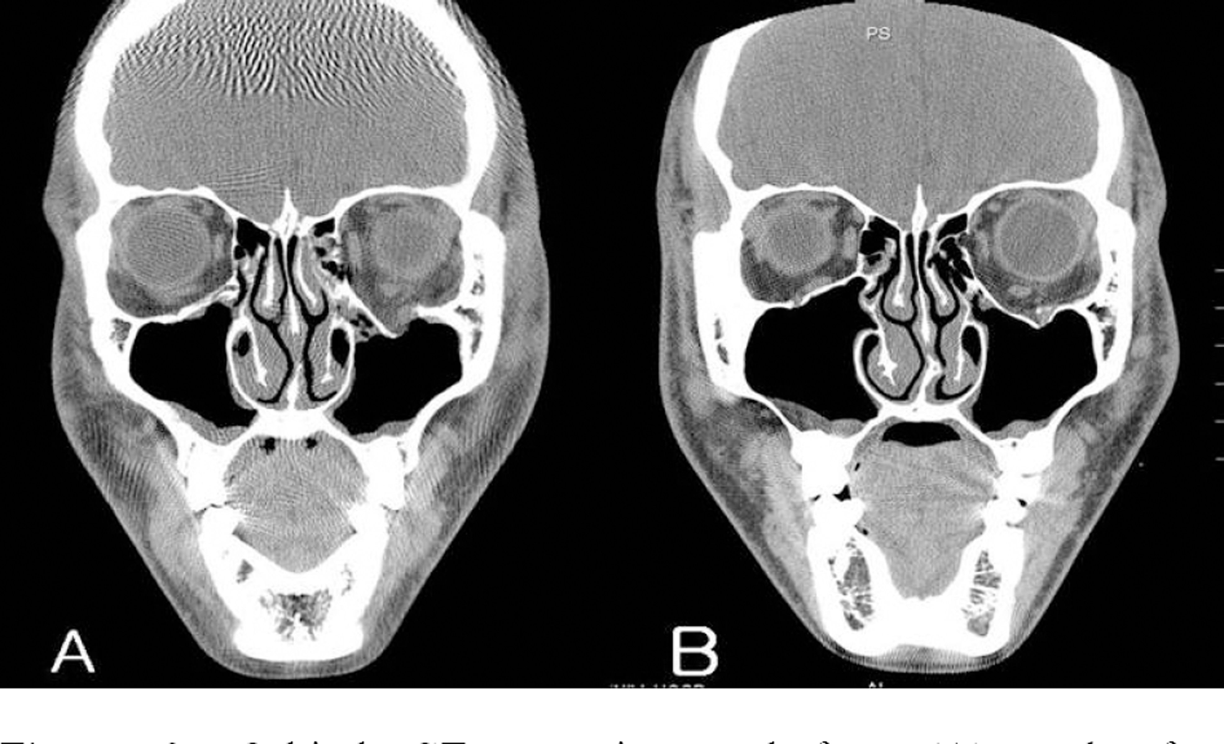
Figure 2. Orbital CT scanning : before (A) and after operation (B).

Figure 3. Measurement of orbital volume by Rapidia 2.8 program: before (A) and after operation (B).
Cited by 2 articles
-
Long-Term Results of Reconstruction of Orbital Wall Fracture With Resorbable Copolymer Mesh
Jae Seok Im, Do Hoon Park, Ju Young Kwak
J Korean Ophthalmol Soc. 2009;50(7):976-983. doi: 10.3341/jkos.2009.50.7.976.A Case of Delayed Orbital Cellulitis after Orbital Wall Fracture Repair Using Absorbable Implant
Jinsoo Kim, Min Joung Lee
J Korean Ophthalmol Soc. 2016;57(7):1165-1169. doi: 10.3341/jkos.2016.57.7.1165.
Reference
-
References
1. Rubin PA, Bilyk JP, Shore JW. Management of orbital trauma: fractures, hemorrhage, and traumatic optic neuropathy. Am Acad Ophthalmol Focal Points. 1994; 7:1–8.2. Dutton JJ. Management of blow-out fractures of the orbital floor. Surv Ophthalmol. 1991; 35:279–80.3. Chi MJ, Jeung JW, Lee JH. Reconstruction of orbital wall fracture with resorbable copolymer mesh. J Korean Ophthalmol Soc. 2006; 47:1021–1030.4. Al-Sukhun J, Törnwall J, Lindqvist C, Konito R. Bioresorbable poly-L/DL-lactide plates are reliable for repairing large inferior orbital wall bony defects: a pilot study. J Oral Maxillofac Surg. 2006; 64:47–55.5. Ploder O, Oeckher M, Klug C. . Follow-up study of treatment of orbital floor fractures: relation of clinical data and software-based CT-analysis. Int J Oral Maxillofac Surg. 2003; 32:257–262.
Article6. Ye J, Kook KH, Lee SY. Evaluation of computer-based volume measurement and porous polyethylene channel implants in reconstruction of large orbital wall fractures. Invest Ophthalmol Vis Sci. 2006; 47:509–513.
Article7. Raskin EM, Millman AL, Lubkin V. . Prediction of late enophthalmos by volumetric analysis of orbital fractures. Ophthal Plast Reconstr Surg. 1998; 14:19–26.
Article8. Fan X, Li J, Li H. . Computer-assisted orbital volume measurement in the surgical correction of late enophthalmos caused by blowout fractures. Ophthal Plast Reconstr Surg. 2003; 19:207–211.
Article9. Mathog RH. Management of orbital blow-out fractures. Otolaryngol Clin North Am. 1991; 24:79–91.
Article10. Choi JC, Fleming JC, Aitken PA, Shore JW. Porous polyethylene channel implants: a modified porous polyethylene sheet implant designed for repairs of large and complex orbital wall fractures. Ophthal Plast Reconstr Surg. 1999; 15:56–66.11. Al-Sukhun J, Lindqvist C. A comparative study of 2 implants used to repair inferior orbital wall bony defects: autogenous bone graft versus bioresorbable poly-L/DL-lactide plate. J Oral Maxillofac Surg. 2006; 64:1038–1048.12. Jeon SY, Kwon JH, Kim JP. . Endoscopic intranasal reduction of the orbit in isolated blowout fractures. Acta OtoLaryngol. 2007; 558:S102–9.
Article13. Putterman AM. Management of blow out fractures of the orbital floor. The conservative approach. Surv Ophthalmol. 1991; 35:292–8.14. Hawes MJ, Dortzbach RK. Surgery on orbital floor fractures. Influence of time of repair and fracture size. Ophthalmology. 1983; 90:1066–70.15. Hosal BM, Beatty RL. Diplopia and enophthalmos after surgical repair of blowout fracture. Orbit. 2002; 21:27–33.
Article16. Yang PJ, Chi NC, Choi GJ. Comparison of surgical outcome between early and delayed repair of orbital wall fracture. Korean Ophthalmol Soc. 2003; 44:1278–84.17. De Man K, Wijngaarde R, Hes J, de Jong PT. Influence of age on the management of blow-out fractures of the orbital floor. Int J Oral Maxillofac Surg. 1991; l20:330–6.
Article18. Harris GJ, Garcia GH, Logani SC. . Orbital blow-out fractures: Correlation of preoperative computed tomography and postoperative ocular motility. Trans Am Ophthalmol Soc. 1998; 96:329–47.
- Full Text Links
-
- Actions
-
Cited
- CITED
-
- Close
- Share
-
- Similar articles
-
- Results of Reconstruction of Orbital Wall Fracture With Bioresorbable Plate
- Anatomical Reconstruction of the Medial Orbital Wall Fracture
- Quantitative Analysis of the Orbital Volume Change in Isolated Zygoma Fracture
- The Normal Value of Adult Korean Orbital Volume in Three-Dimensional Computerized Tomography
- Orbital Wall Reconstruction with Osteoconductive Unsintered Hydroxyapatite/Poly L-Lactide
