Korean J Clin Microbiol.
2010 Sep;13(3):135-139. 10.5145/KJCM.2010.13.3.135.
Fungemia due to Exophiala dermatitidis
- Affiliations
-
- 1Department of Laboratory Medicine, Chonnam National University Medical School, Gwangju, Korea. shinjh@chonnam.ac.kr
- KMID: 1461629
- DOI: http://doi.org/10.5145/KJCM.2010.13.3.135
Abstract
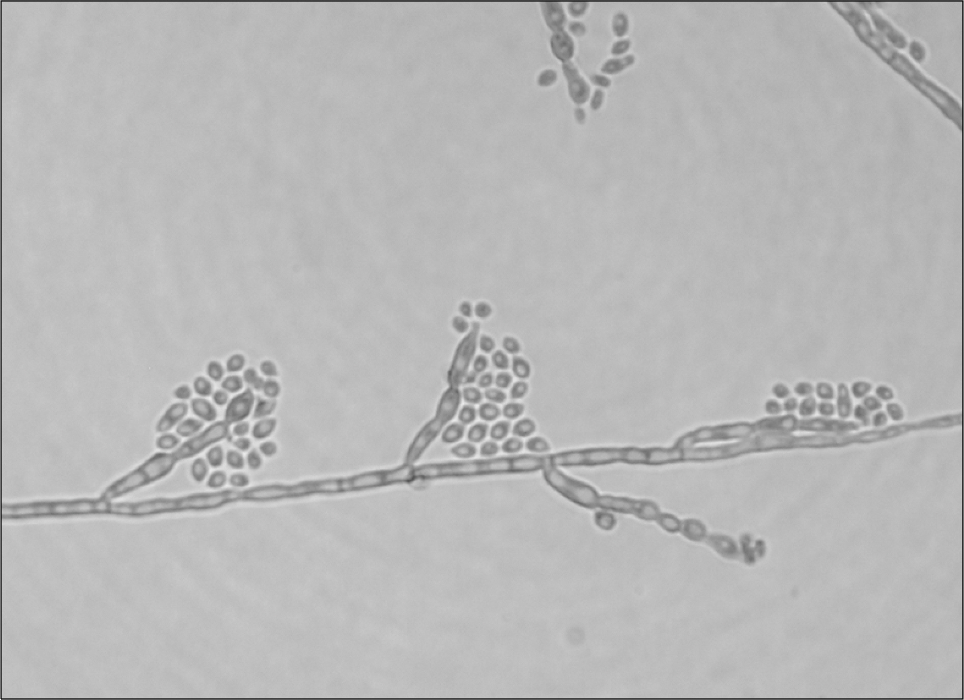
- We report a rare case of fungemia due to Exophiala (Wangiella) dermatitidis in a 4-month-old female infant who was admitted to an intensive care unit with sudden infant death syndrome (SIDS). E. dermatitidiswas repeatedly isolated from blood cultures (on the 28th and 32nd day of hospitalization) of the patient, who died on the 44th day of hospitalization. The fungus was identified by its morphological characteristics and DNA sequencing of both the D1/D2 domain and the ITS region of rDNA. To our knowledge, this is the first reported case of E. dermatitidis fungemia in Korea.
Keyword
MeSH Terms
Figure
Reference
-
1. Nachman S, Alpan O, Malowitz R, Spitzer ED. Catheter-associated fungemia due to Wangiella (Exophiala) dermatitidis. J Clin Microbiol. 1996; 34:1011–3.
Article2. Hohl PE, Holley HP Jr, Prevost E, Ajello L, Padhye AA. Infections due to Wangiella dermatitidis in humans: report of the first documented case from the United States and a review of the literature. Rev Infect Dis. 1983; 5:854–64.
Article3. Vlassopoulos D, Kouppari G, Arvanitis D, Papaefstathiou K, Dounavis A, Velegraki A, et al. Wangiella dermatitidis peritonitis in a CAPD patient. Perit Dial Int. 2001; 21:96–7.
Article4. Chang CL, Kim DS, Park DJ, Kim HJ, Lee CH, Shin JH. Acute cerebral phaeohyphomycosis due to Wangiella dermatitidis accompanied by cerebrospinal fluid eosinophilia. J Clin Microbiol. 2000; 38:1965–6.5. Kim DS, Yoon YM, Kim SW. Phaeohyphomycosis due to Exophiala dermatitidis successfully treated with itraconazole. Korean J Med Mycol. 1999; 4:79–83.6. Hong KH, Kim JW, Jang SJ, Yu E, Kim EC. Liver cirrhosis caused by Exophiala dermatitidis. J Med Microbiol. 2009; 58:674–7.
Article7. Sugita T, Nishikawa A, Ikeda R, Shinoda T. Identification of medically relevant Trichosporon species based on sequences of internal transcribed spacer regions and construction of a database for Trichosporon identification. J Clin Microbiol. 1999; 37:1985–93.8. Kurtzman CP and Robnett CJ. Identification of clinically important ascomycetous yeasts based on nucleotide divergence in the 5' end of the large-subunit (26S) ribosomal DNA gene. J Clin Microbiol. 1997; 35:1216–23.
Article9. Untereiner WA. Fruiting studies in species of Capronia (Herpo- trichiellaceae). Antonie Van Leeuwenhoek. 1995; 68:3–17.10. Al-Obaid I, Ahmad S, Khan ZU, Dinesh B, Hejab HM. Catheter-associated fungemia due to Exophiala oligosperma in a leukemic child and review of fungemia cases caused by Exophiala species. Eur J Clin Microbiol Infect Dis. 2006; 25:729–32.
Article11. Lim WH, Shin JH, Suh SP, Ryang DW. Diagnosis of central venous catheter-related sepsis using differential quantitative blood cultures. Korean J Clin Pathol. 1998; 18:208–14.12. de Hoog GS, Matos T, Sudhadham M, Luijsterburg KF, Haase G. Intestinal prevalence of the neurotropic black yeast Exophiala (Wangiella) dermatitidis in healthy and impaired individuals. Mycoses. 2005; 48:142–5.
Article13. Woollons A, Darley CR, Pandian S, Arnstein P, Blackee J, Paul J. Phaeohyphomycosis caused by Exophiala dermatitidis following intra-articular steroid injection. Br J Dermatol. 1996; 135:475–7.
Article14. de Hoog GS and Vitale RG. Bipolaris, Exophiala, Scedosporium, Sporothriz, and Other Dematiaceous Fungi. Murray PR, Baron EJ, Jeorgensen JH, Landry ML, Pfaller MA, editors. Manual of Clinical Microbiology. 9th ed.Washington D.C.: ASM Press;2006. p. 1898–917.15. Petti CA. Detection and identification of microorganisms by gene amplification and sequencing. Clin Infect Dis. 2007; 44:1108–14.16. Vitale RG, De Hoog GS, Verweij PE. In vitro activity of amphotericin B, itraconazole, terbinafine and 5-fluocytosine against Exophiala spinifera and evaluation of post-antifungal effects. Med Mycol. 2003; 41:301–7.
Article
- Full Text Links
- Actions
-
Cited
- CITED
-
- Close
- Share
- Similar articles
-
- Phaeohyphomycosis Due to Exophiala dermatitidis Successfully Treated with Itraconazole
- Molecular Phylogenetics of Exophiala Species Isolated from Korea
- A Case of Phaeomycotic Subcutaneous Abscess Caused by Wangiella Dermatitidis
- Molecular Analysis of Exophiala Species Using Molecular Markers
- Antifungal Susceptibility Testing of Dematiaceous Fungi Using Etest


