J Cardiovasc Ultrasound.
2010 Mar;18(1):6-11. 10.4250/jcu.2010.18.1.6.
Usefulness of Mitral Annulus Velocity for the Early Detection of Left Ventricular Dysfunction in a Rat Model of Diabetic Cardiomyopathy
- Affiliations
-
- 1Cardiovascular Center, Seoul National University Hospital, Department of Internal Medicine, Seoul National University College of Medicine, Seoul, Korea. kimdamas@snu.ac.kr
- KMID: 1457198
- DOI: http://doi.org/10.4250/jcu.2010.18.1.6
Abstract
- BACKGROUND
Diabetic cardiomyopathy (DMCMP) is characterized by myocardial dysfunction regardless of coronary artery disease in diabetic patients. The features of LV dysfunction in rat model of type 1 DM induced by streptozocin, are variable and controversial. Thus, we tested the usefulness of tissue Doppler imaging in the early detection of ventricular dysfunction in a rat model of DMCMP. METHODS: Diabetes was induced by intra-peritoneal injection of streptozocin (70 mg/kg) in 8 weeks of Sprague-Dawley rat. Diagnosis of diabetes was defined as venous glucose level over 350 mg/dL 48 hrs after streptozocin injection. Echocardiography was done at baseline and 10 weeks after diabetes induction both in diabetes group (n=15) and normal control (n=10). After echocardiography at 10 weeks, invasive hemodynamic measurement using miniaturized conductance catheter was done in both groups. RESULTS: Ten weeks after diabetes induction, heart and lung mass indexes of diabetes were larger than those of normal control (3.2+/-0.3 vs. 2.4+/-0.2 mg/g, p<0.001, 5.5+/-1.1 vs. 3.6+/-0.6 mg/g, p<0.001, respectively). In echocardiographic data, s' (2.4+/-0.4 vs. 3.1+/-0.5 cm/s, p<0.001), e' velocity of mitral annulus (2.9+/-0.6 vs. 3.8+/-1.1 cm/s, p<0.001), and E/e' ratio (27.1+/-5.6 vs. 19.7+/-2.6, p<0.001) were impaired in diabetes group. In hemodynamic measurement, there were no differences in ejection fraction, peak dP/dt between the diabetic group and normal control. However, load independent indexes of contractility, the slope of the end-systolic pressure volume relation (0.18+/-0.07 vs. 0.62+/-0.18 mmHg/microL, p<0.001) and preload recruitable stroke work (51.8+/-22.0 vs. 90.9+/-22.5 mmHg, p<0.001) were impaired in diabetic group compared to normal control. CONCLUSION: In a rat model of diabetic cardiomyopathy, tissue Doppler imaging of mitral annulus can be a good modality for early detection of myocardial dysfunction.
MeSH Terms
Figure
-
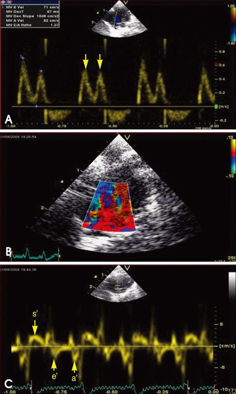
Fig. 1 Mitral inflow (A), tissue Doppler imaging of mitral annulus (B), mitral annulus velocity, s', e' and a', respectively (C) (arrow).
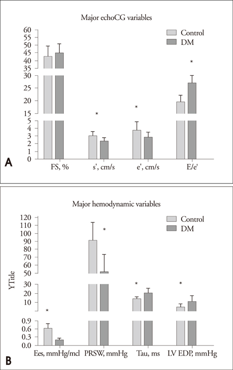
Fig. 2 A: Major echocardiographic variables. B: Bar graphs demonstrate major hemodynamic parameters including Ees, PRSW, tau and LV EDP. *p<0.05 for difference from control. FS: fractional shortening, s': mitral annulus peak systolic velocity, e': mitral annulus early diastolic velocity, a': mitral annulus later diastolic velocity.
Cited by 1 articles
-
Pharmacodynamic Analysis of the Influence of Propofol on Left Ventricular Long-Axis Systolic Performance in Cardiac Surgical Patients
Ji-Yeon Bang, Sooyoung Kim, Byung-Moon Choi, Tae-Yop Kim
J Korean Med Sci. 2019;34(16):. doi: 10.3346/jkms.2019.34.e132.
Reference
-
1. Marwick TH. Diabetic heart disease. Heart. 2006. 92:296–300.
Article2. Rubler S, Dlugash J, Yuceoglu YZ, Kumral T, Branwood AW, Grishman A. New type of cardiomyopathy associated with diabetic glomerulosclerosis. Am J Cardiol. 1972. 30:595–602.
Article3. Boudina S, Abel ED. Diabetic cardiomyopathy revisited. Circulation. 2007. 115:3213–3223.
Article4. Zarich SW, Nesto RW. Diabetic cardiomyopathy. Am Heart J. 1989. 118:1000–1012.
Article5. Cai L, Kang YJ. Oxidative stress and diabetic cardiomyopathy: a brief review. Cardiovasc Toxicol. 2001. 1:181–193.
Article6. Fiordaliso F, Li B, Latini R, Sonnenblick EH, Anversa P, Leri A, Kajstura J. Myocyte death in streptozotocin-induced diabetes in rats in angiotensin II- dependent. Lab Invest. 2000. 80:513–527.
Article7. Tschöpe C, Walther T, Königer J, Spillmann F, Westermann D, Escher F, Pauschinger M, Pesquero JB, Bader M, Schultheiss HP, Noutsias M. Prevention of cardiac fibrosis and left ventricular dysfunction in diabetic cardiomyopathy in rats by transgenic expression of the human tissue kallikrein gene. FASEB J. 2004. 18:828–835.
Article8. Tschöpe C, Spillmann F, Rehfeld U, Koch M, Westermann D, Altmann C, Dendorfer A, Walther T, Bader M, Paul M, Schultheiss HP, Vetter R. Improvement of defective sarcoplasmic reticulum Ca2+ transport in diabetic heart of transgenic rats expressing the human kallikrein-1 gene. FASEB J. 2004. 18:1967–1969.
Article9. Westermann D, Rutschow S, Jäger S, Linderer A, Anker S, Riad A, Unger T, Schultheiss HP, Pauschinger M, Tschöpe C. Contributions of inflammation and cardiac matrix metalloproteinase activity to cardiac failure in diabetic cardiomyopathy: the role of angiotensin type 1 receptor antagonism. Diabetes. 2007. 56:641–646.
Article10. Boyer JK, Thanigaraj S, Schechtman KB, Pérez JE. Prevalence of ventricular diastolic dysfunction in asymptomatic, normotensive patients with diabetes mellitus. Am J Cardiol. 2004. 93:870–875.
Article11. Fang ZY, Yuda S, Anderson V, Short L, Case C, Marwick TH. Echocardiographic detection of early diabetic myocardial disease. J Am Coll Cardiol. 2003. 41:611–617.
Article12. Candido R, Forbes JM, Thomas MC, Thallas V, Dean RG, Burns WC, Tikellis C, Ritchie RH, Twigg SM, Cooper ME, Burrell LM. A breaker of advanced glycation end products attenuates diabetes-induced myocardial structural changes. Circ Res. 2003. 92:785–792.
Article13. Tschöpe C, Walther T, Yu M, Reinecke A, Koch M, Seligmann C, Heringer SB, Pesquero JB, Bader M, Schultheiss H, Unger T. Myocardial expression of rat bradykinin receptors and two tissue kallikrein genes in experimental diabetes. Immunopharmacology. 1999. 44:35–42.
Article14. Georgakopoulos D, Mitzner WA, Chen CH, Byrne BJ, Millar HD, Hare JM, Kass DA. In vivo murine left ventricular pressure-volume relations by miniaturized conductance micromanometry. Am J Physiol. 1998. 274:H1416–H1422.15. Pacher P, Nagayama T, Mukhopadhyay P, Bátkai S, Kass DA. Measurement of cardiac function using pressure-volume conductance catheter technique in mice and rats. Nat Protoc. 2008. 3:1422–1434.
Article16. Van Linthout S, Riad A, Dhayat N, Spillmann F, Du J, Dhayat S, Westermann D, Hilfiker-Kleiner D, Noutsias M, Laufs U, Schultheiss HP, Tschöpe C. Anti-inflammatory effects of atorvastatin improve left ventricular function in experimental diabetic cardiomyopathy. Diabetologia. 2007. 50:1977–1986.
Article17. Westermann D, Van Linthout S, Dhayat S, Dhayat N, Escher F, Bücker-Gärtne C, Spillmann F, Noutsias M, Riad A, Schultheiss HP, Tschöpe C. Cardioprotective and anti-inflammatory effects of interleukin converting enzyme inhibition in experimental diabetic cardiomyopathy. Diabetes. 2007. 56:1834–1841.
Article18. Yoon YS, Uchida S, Masuo O, Cejna M, Park JS, Gwon HC, Kirchmair R, Bahlman F, Walter D, Curry C, Hanley A, Isner JM, Losordo DW. Progressive attenuation of myocardial vascular endothelial growth factor expression is a seminal event in diabetic cardiomyopathy: restoration of microvascular homeostasis and recovery of cardiac function in diabetic cardiomyopathy after replenishment of local vascular endothelial growth factor. Circulation. 2005. 111:2073–2085.
Article19. Nagueh SF. Tissue Doppler imaging for the assessment of left ventricular diastolic function. J Cardiovasc Ultrasound. 2008. 16:76–79.
Article20. Ommen SR, Nishimura RA, Appleton CP, Miller FA, Oh JK, Redfield MM, Tajik AJ. Clinical utility of Doppler echocardiography and tissue Doppler imaging in the estimation of left ventricular filling pressures: A comparative simultaneous Doppler-catheterization study. Circulation. 2000. 102:1788–1794.
Article21. Prunier F, Gaertner R, Louedec L, Michel JB, Mercadier JJ, Escoubet B. Doppler echocardiographic estimation of left ventricular end-diastolic pressure after MI in rats. Am J Physiol Heart Circ Physiol. 2002. 283:H346–H352.22. Kim YJ. Doppler Tissue Imaging. J Korean Soc Echocardiogr. 2003. 11:63–69.
Article
- Full Text Links
-
- Actions
-
Cited
- CITED
-
- Close
- Share
-
- Similar articles
-
- Assessment of Diastolic Function Using Mitral Annulus Velocity by Doppler Tissue Velocity in the Patients with Left Ventricular Hypertrophy
- A Comparative Study of Left Ventricular Diastolic Function between Hypertrophic Cardiomyopathy and Hypertensive Patients with Left Ventricular Hypertrophy
- Assessment of Normal Mitral Annulus Velocity by Doppler Tissue Imaging
- Assessment of Left Ventricular Diastolic Dysfunction by Intraventricular Dispersion of Early Diastolic Filling
- Assessment of Diastolic Function using Mitral Annulus Velocity by Doppler Tissue Velocity in the Patients with Hypertension
