J Korean Soc Radiol.
2011 Aug;65(2):171-179. 10.3348/jksr.2011.65.2.171.
Imaging Spectrums of the Male Breast Diseases: A Pictorial Essay
- Affiliations
-
- 1Department of Radiology, Kangnam Sacred Heart Hospital, Hallym University College of Medicine, Seoul, Korea.
- 2Department of Radiology, Sungkyunkwan University College of Medicine, Kangbuk Samsung Hospital, Seoul, Korea. dr_philic@naver.com
- 3Department of Pathology, Kangnam Sacred Heart Hospital, Hallym University College of Medicine, Seoul, Korea.
- KMID: 1443482
- DOI: http://doi.org/10.3348/jksr.2011.65.2.171
Abstract
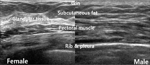
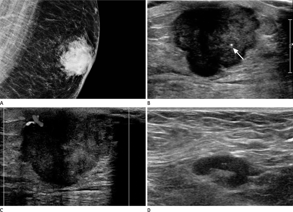
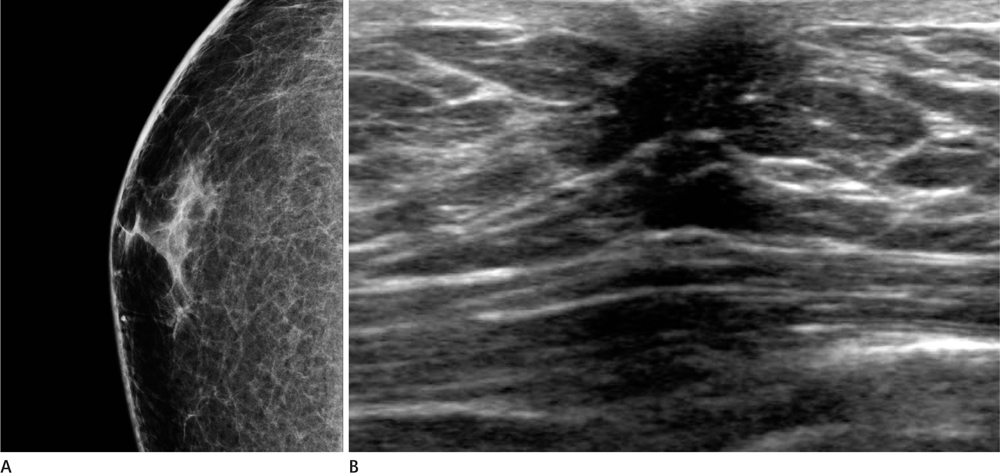
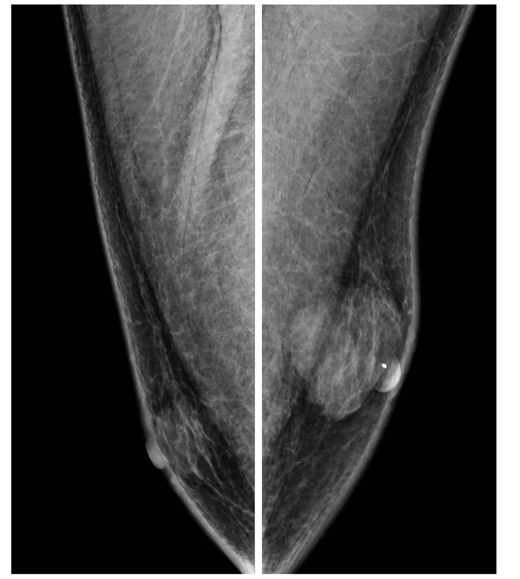
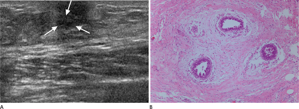
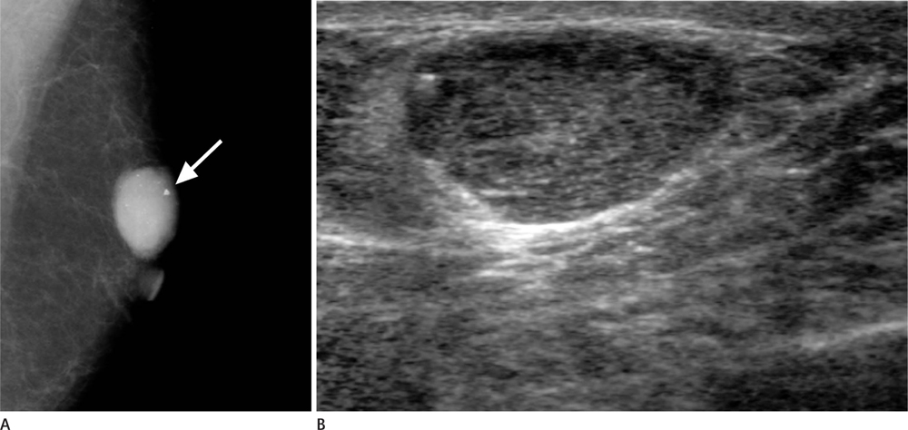
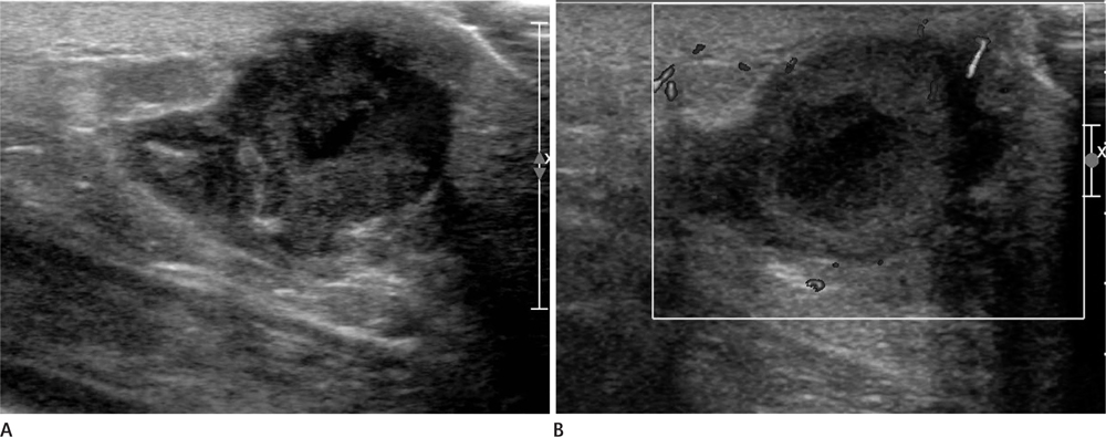
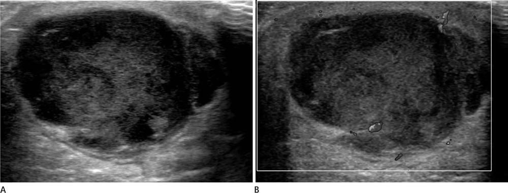
- Most described male breast lesions, such as gynecomastia, are benign. The overall incidence of male breast cancer is less than 3%. Like women, common presentations of male breast diseases are palpable lumps or tenderness. Physical examination, mammography and ultrasound are generally used for work-up of breast diseases in both women and men. However, men do not undergo screening mammograms; all male patients are examined in symptomatic cases only. Therefore, all male breast examinations are diagnostic, whereas the majority of the examinations for women are for screening purpose. The differentiation between benign and malignant breast lesions is important, especially for men, because the reported prognosis of male breast cancer is poor due to delayed diagnosis. In this article, we review the spectrum of male breast diseases, from benign to malignant, and illustrate their ultrasonographic and mammographic imaging features.
MeSH Terms
Figure
Reference
-
1. Appelbaum AH, Evans GF, Levy KR, Amirkhan RH, Schumpert TD. Mammographic appearances of male breast disease. Radiographics. 1999; 19:559–568.2. Chen L, Chantra PK, Larsen LH, Barton P, Rohitopakarn M, Zhu EQ, et al. Imaging characteristics of malignant lesions of the male breast. Radiographics. 2006; 26:993–1006.3. Günhan-Bilgen I, Bozkaya H, Ustün EE, Memiş A. Male breast disease: clinical, mammographic, and ultrasonographic features. Eur J Radiol. 2002; 43:246–255.4. Wise GJ, Roorda AK, Kalter R. Male breast disease. J Am Coll Surg. 2005; 200:255–269.5. Shetty MK, Shah YP. Sonographic findings in focal fibrocystic changes of the breast. Ultrasound Q. 2002; 18:35–40.6. Gateley CA. Male breast disease. The Breast. 1998; 7:121–127.7. Kook SH, Kim MS, Pae WK. Atypical sonographic patterns of fibroadenoma of the breast: pathologic correlation. J Korean Radiol Soc. 1999; 40:597–602.8. Reddy KM, Meyer CE, Nakdjevani A, Shrotria S. Idiopathic granulomatous mastitis in the male breast. Breast J. 2005; 11:73.9. Hovanessian Larsen LJ, Peyvandi B, Klipfel N, Grant E, Iyengar G. Granulomatous lobular mastitis: imaging, diagnosis, and treatment. AJR Am J Roentgenol. 2009; 193:574–581.10. Vourtsi A, Zervoudis S, Pafiti A, Athanasiadis S. Male breast hemangioma--a rare entity: a case report and review of the literature. Breast J. 2006; 12:260–262.
- Full Text Links
- Actions
-
Cited
- CITED
-
- Close
- Share
- Similar articles
-
- Sonographic Features of Palpable Breast and Axillary Lesions in Adult Male Patients: A Pictorial Essay
- Breast lesions during pregnancy and lactation: a pictorial essay
- Imaging Features of Inflammatory Breast Disorders: A Pictorial Essay
- Imaging Spectrum of Augmented Breast and Post-Mastectomy Reconstructed Breast with Common Complications: A Pictorial Essay
- Extra-Mammary Findings Detected on Breast Magnetic Resonance Imaging: A Pictorial Essay

