J Korean Soc Radiol.
2012 Mar;66(3):275-277. 10.3348/jksr.2012.66.3.275.
Extra-Gastrointestinal Stromal Tumor of Retroperitoneal Origin: A Case Report
- Affiliations
-
- 1Department of Radiology, Gachon University of Medicine and Science, Gil Medical Center, Incheon, Korea. hskim@gilhospital.com
- KMID: 1439426
- DOI: http://doi.org/10.3348/jksr.2012.66.3.275
Abstract
- Extragastrointestinal stromal tumors (EGIST) are relatively rare, and cases originating in the retroperitoneum even rarer. We report a 60-year-old woman who presented with an EGIST originating in the retroperitoneum. Computed tomography results demonstrated a soft tissue mass on the right side of the retroperitoneum. The tumor abutted the duodenum, head of the pancreas, and right kidney. The mass was surgically proven to be a retroperitoneal tumor and histopathologically proven to be a retroperitoneal EGIST.
Figure
-
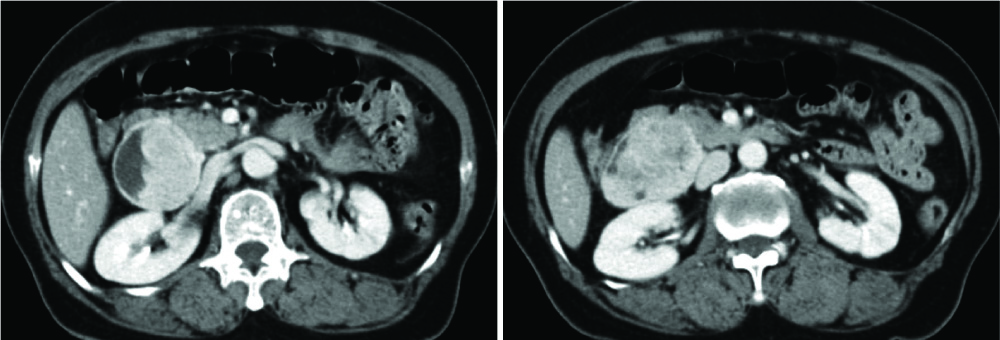
Fig. 1 A 60-year-old woman with a 6.5 × 5.0 × 6.0 cm retroperitoneal mass. Post-contrast CT scans shows well circumscribed, lobulated and heterogeneous enhanced solid and cystic mass arising from retroperitoneum.
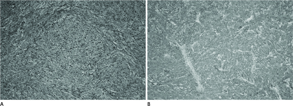
Fig. 2 A 60-year-old woman with a primary retroperitoneal gastrointestinal stromal tumor. A. Microscopic findings of primary retroperitoneal gastrointestinal stromal tumor. The tumor is composed of spindle shaped cells arranged in fascicular pattern (H&E staining, × 100). B. Diffuse strong positive staining for c-kit protein is observed in immunochemistry test (× 100).
Reference
-
1. Seo MH, Shim JC, Joo M, Ryu SJ, Lee GJ, Kim HK. Malignant gastrointestinal stromal tumor of mesentery origin: case report. J Korean Radiol Soc. 2002; 47:631–634.2. Ko ES, Bae K, Jeon KN, Kim JS, You JJ, Ryeom HK, et al. Extragastrointestinal stromal tumor presenting a large multilocular cystic mass arising from the greater omentum: a case report. J Korean Radiol Soc. 2004; 51:533–536.3. Miettinen M, Monihan JM, Sarlomo-Rikala M, Kovatich AJ, Carr NJ, Emory TS, et al. Gastrointestinal stromal tumors/smooth muscle tumors (GISTs) primary in the omentum and mesentery: clinicopathologic and immunohistochemical study of 26 cases. Am J Surg Pathol. 1999; 23:1109–1118.4. Reith JD, Goldblum JR, Lyles RH, Weiss SW. Extragastrointestinal (soft tissue) stromal tumors: an analysis of 48 cases with emphasis on histologic predictors of outcome. Mod Pathol. 2000; 13:577–585.5. Miettinen M, Sarlomo-Rikala M, Lasota J. Gastrointestinal stromal tumors: recent advances in understanding of their biology. Hum Pathol. 1999; 30:1213–1220.6. Fletcher CD, Berman JJ, Corless C, Gorstein F, Lasota J, Longley BJ, et al. Diagnosis of gastrointestinal stromal tumors: a consensus approach. Hum Pathol. 2002; 33:459–465.7. Kim HC, Lee JM, Kim SH, Kim KW, Lee M, Kim YJ, et al. Primary gastrointestinal stromal tumors in the omentum and mesentery: CT findings and pathologic correlations. AJR Am J Roentgenol. 2004; 182:1463–1467.
- Full Text Links
-
- Actions
-
Cited
- CITED
-
- Close
- Share
-
- Similar articles
-
- Primary Retroperitoneal Malignant Gastrointestinal Stromal Tumor Mimicking Adrenal Mass
- Diagnosis of a Gastrointestinal Stromal Tumor Presenting as a Prostatic Mass: A Case Report
- Carney Triad in an Adult with Aggressive Behavior: The First Case in Korea
- Malignant Gastrointestinal Stromal Tumor of Mesentery Origin: Case Report
- Hypertensive crisis during wide excision of gastrointestinal stromal cell tumor (GIST): Undiagnosed paraganglioma: A case report
