Primary Retroperitoneal Malignant Gastrointestinal Stromal Tumor Mimicking Adrenal Mass
- Affiliations
-
- 1Department of Surgery, Seoul National University Boramae Hospital, Seoul, Korea. jkchung@brm.co.kr
- 2Department of Pathology, Seoul National University Boramae Hospital, Seoul, Korea.
- KMID: 1750706
- DOI: http://doi.org/10.4174/jkss.2009.77.5.357
Abstract
- Gastrointestinal stromal tumor (GIST) is the most common non-epithelial tumor in the gastrointestinal tract. Although GIST occurs mainly in the gastrointestinal tract, it also occurs, rarely, in non-gastrointestinal tract and in this case, it is often named as extra-gastrointestinal stromal tumor (EGIST). We experienced a 68-year-old male patient who had been diagnosed preoperatively with accidentaloma of the left adrenal gland by computed tomography, and finally diagnosed as primary retroperitoneal malignant GIST, postoperatively. The operation was performed via anterior abdominal approach, and complete surgical resection was done for a 7 cm sized retroperitoneal tumor near the left adrenal gland. Primary retroperitoneal malignant GIST was the final pathologic diagnosis and the size of the tumor was 6.5 cm and the mitotic count was 7 per high-power field. Diffuse strong positive staining for c-kit protein, CD34 and negative staining for desmin were observed in a immunohistochemistry test. We report here the unusual case of primary retroperitoneal malignant GIST mimicking adrenal mass.
Keyword
MeSH Terms
Figure
-
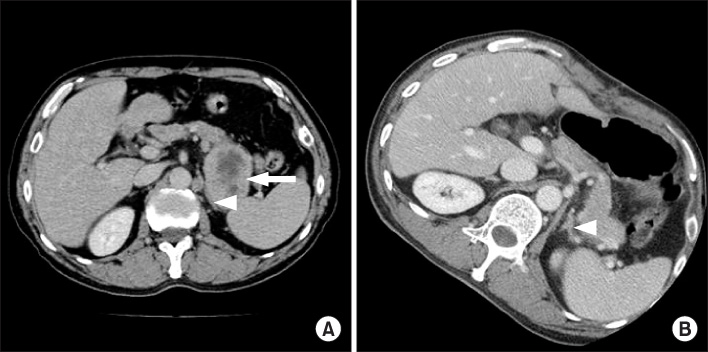
Fig. 1 (A) Well-marginated and well enhancing mass with central necrosis is found (arrow) near lateral limb of left adrenal gland (arrow head) and size of tumor is about 6.2×4.6 cm in preoperative computerized tomography. (B) Tumor is no longer found and normal appearance of lateral limb of left adrenal gland is found (arrow head) in postoperative computerized tomography.

Fig. 2 Well-developed membranous capsular tissue is shown in gross specimen of primary retroperitoneal malignant gastrointestinal tumor (A) and solid tumor with gray-white flesh tissue content is shown at cut surface (B).

Fig. 3 (A) Microscopic findings of primary retroperitoneal malignant gastrointestinal tumor (H&E staining, ×100). (B) Mitosis is observed in center of field (circle) (H&E staining, ×400). Diffuse strong positive staining for c-kit protein (C) and CD34 (D) is observed in immunohistochemistry test (×100).
Cited by 1 articles
-
A Case of Primary Extragastrointestinal Stromal Tumor Presenting as Peritoneal Dissemination
Hong Jun Yang, Tae Ho Kim, Min Kyoung Park, Chang Hoon Lim, Kee Hyun Lee, Chang Whan Kim, Sok Won Han, Jean A Kim
Korean J Gastroenterol. 2010;56(5):319-323. doi: 10.4166/kjg.2010.56.5.319.
Reference
-
1. Fletcher CD, Berman JJ, Corless C, Gorstein F, Lasota J, Longley BJ, et al. Diagnosis of gastrointestinal stromal tumors: a consensus approach. Hum Pathol. 2002. 33:459–465.2. Park SW, Lee W, Huh GY, Chung MK. Gastrointestinal stromal tumor of prostate. Korean J Urol. 2008. 49:383–385.3. Kim ST, Paik KY, Chung JC, Choi DW. Extra-Gastrointestinal stromal tumor of the pancreas with c-KIT gene mutation: Report of a case. Korean J Hepatobiliary Pancreat Surg. 2007. 11:63–66.4. Yoon SJ, Lee JH, Kim HA, Cheon SH, Lee JH, Lee RA, et al. Recurrence patterns and clinical behavior of gastrointestinal stromal tumors (GISTs). J Korean Surg Soc. 2006. 70:430–436.5. Reith JD, Goldblum JR, Lyles RH, Weiss SW. Extragastrointestinal (soft tissue) stromal tumors: an analysis of 48 cases with emphasis on histologic predictors of outcome. Mod Pathol. 2000. 13:577–585.6. Miettinen M, Monihan JM, Sarlomo-Rikala M, Kovatich AJ, Carr NJ, Emory TS, et al. Gastrointestinal stromal tumors/smooth muscle tumors (GISTs) primary in the omentum and mesentery: clinicopathologic and immunohistochemical study of 26 cases. Am J Surg Pathol. 1999. 23:1109–1118.7. Yeung CK, Yuen CH, Chan IK, Chu RW. Malignant extra-gastrointestinal stromal tumour of diaphragm. ANZ J Surg. 2008. 78:923–924.8. Nagase S, Mikami Y, Moriya T, Niikura H, Yoshinaga K, Takano T, et al. Vaginal tumors with histologic and immunocytochemical feature of gastrointestinal stromal tumor: two cases and review of the literature. Int J Gynecol Cancer. 2007. 17:928–933.9. Miettinen M, Lasota J. Gastrointestinal stromal tumors--definition, clinical, histological, immunohistochemical, and molecular genetic features and differential diagnosis. Virchows Arch. 2001. 438:1–12.10. Blay JY, Bonvalot S, Casali P, Choi H, Debiec-Richter M, Dei Tos AP, et al. Consensus meeting for the management of gastrointestinal stromal tumors. Report of the GIST Consensus Conference of 20~21 March 2004, under the auspices of ESMO. Ann Oncol. 2005. 16:566–578.
- Full Text Links
-
- Actions
-
Cited
- CITED
-
- Close
- Share
-
- Similar articles
-
- A Case of Incidentally Detected Silent Adrenal Pheochromocytoma, Mimicking as an Intraperitoneal Tumor With Unusual Computed Tomography Findings
- Carney Triad in an Adult with Aggressive Behavior: The First Case in Korea
- Mature Teratoma in the Adrenal Gland
- Adrenal Tuberculosis Mimicking a Malignant Tumor with Primary Adrenal Insufficiency
- Retroperitoneal Tumors Mimicking Adrenal Tumor
