Displacement of dental implants into the focal osteoporotic bone marrow defect: a report of three cases
- Affiliations
-
- 1Department of Oral and Maxillofacial Surgery, Gachon University Gil Medical Center, Incheon, Korea. umincw@gilhospital.com
- KMID: 1430501
- DOI: http://doi.org/10.5125/jkaoms.2013.39.2.94
Abstract
- Focal osteoporotic bone marrow defect (FOBMD) is a radiolucent area corresponding to the presence of hematopoietic tissue rarely found in the jaws. FOBMD is most commonly located in the mandibular edentulous posterior area of a middle-aged female. From November 2011 to November 2012, we experienced three cases involving removal of implants that had accidentally fallen into the FOBMD area. All patients happened to be female, with a mean age of 54 years (range: 51-60 years). One case involved hypoesthesia of the lower lip and chin, while two cases healed without any complication. Displacement of an implant into the FOBMD area is an unusual event, which occurs rarely during placement of a dental fixture. The purpose of this study was to report on three cases of FOBMD and to provide a review of related literature.
Keyword
MeSH Terms
Figure
-
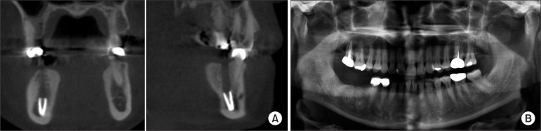
Fig. 1 A. Preoperative coronal (left) and sagittal (right) cone-beam computed tomography showing the displacement of the #46 implant near the mandibular border of case 1. B. Follow-up panorama taken 3 months after the removal operation indicates the resolution of the lesion on the #46 area.
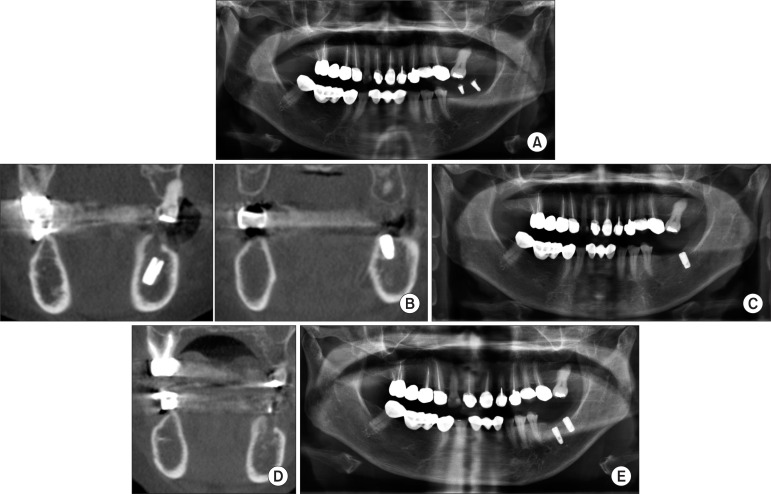
Fig. 2 A. Preoperative panorama showing normal bone on the left mandibular body without any cystic lesion of case 2. B. Preoperative cone-beam computed tomography showing the displacement of #36 implant (left) near the inferior alveolar nerve and normal position of #37 implant (right) of case 2. C. Postoperative panoramic view of #36 implant removal surgery of case 2. D. Follow-up computed tomography taken 3 months after the removal operation indicates the resolution of the lesion of case 2. E. Follow-up panoramic view after the placement of # 36 implant of case 2.
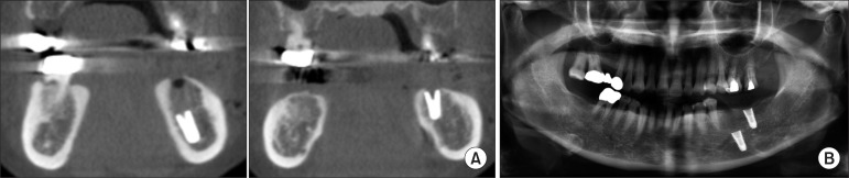
Fig. 3 A. Preoperative cone-beam computed tomography view of the displacement of #36 implant (left) near the mandibular border area and normal bone state of #37 implant (right) of case 3. B. Preoperative panorama showing normal cancellous bone without any cystic lesion of case 3.
Reference
-
1. Schneider LC, Mesa ML, Fraenkel D. Osteoporotic bone marrow defect: radiographic features and pathogenic factors. Oral Surg Oral Med Oral Pathol. 1988; 65:127–129. PMID: 3277106.
Article2. Bravo-Calderón DM, Oliveira DT, Martins Dos Santos WH. Bilateral osteoporotic bone marrow defects of the mandible: a case report. Head Face Med. 2012; 8:22. PMID: 22873712.
Article3. Lipani CS, Natiella JR, Greene GW Jr. The hematopoietic defect of the jaws: a report of sixteen cases. J Oral Pathol. 1982; 11:411–416. PMID: 6819349.
Article4. Shankland WE, Bouquot JE. Focal osteoporotic marrow defect: report of 100 new cases with ultrasonography scans. Cranio. 2004; 22:314–319. PMID: 15532316.5. Bayram B, Alaaddinoglu E. Implant-box mandible: dislocation of an implant into the mandible. J Oral Maxillofac Surg. 2011; 69:498–501. PMID: 21238846.
Article6. Lamas Pelayo J, Peñarrocha Diago M, Martí Bowen E, Peñarrocha Diago M. Intraoperative complications during oral implantology. Med Oral Patol Oral Cir Bucal. 2008; 13:E239–E243. PMID: 18379448.7. Makek M, Lello GE. Focal osteoporotic bone marrow defects of the jaws. J Oral Maxillofac Surg. 1986; 44:268–273. PMID: 3457122.
Article8. Sencimen M, Delilbasi C, Gulses A, Okcu KM, Gunhan O, Varol A. Focal osteoporotic hematopoietic bone marrow defect formation around a dental implant: a case report. Int J Oral Maxillofac Implants. 2011; 26:e1–e4. PMID: 21365033.9. CHI AC. Bone pathology. In : Neville BW, Damm DD, Allen CM, Bouquot JE, editors. Oral and maxillofacial pathology. 3rd ed. St. Louis: Saunders-Elsevier;2009. p. 613–677.10. Xanthinaki AA, Choupis KI, Tosios K, Pagkalos VA, Papanikolaou SI. Traumatic bone cyst of the mandible of possible iatrogenic origin: a case report and brief review of the literature. Head Face Med. 2006; 2:40. PMID: 17096860.
Article11. Bender IB, Seltzer S. Roentgenographic and direct observation of experimental lesions in bone: I. 1961. J Endod. 2003; 29:702–706. PMID: 14651274.12. Vlasiadis KZ, Skouteris CA, Velegrakis GA, Fragouli I, Neratzoulakis JM, Damilakis J, et al. Mandibular radiomorphometric measurements as indicators of possible osteoporosis in postmenopausal women. Maturitas. 2007; 58:226–235. PMID: 17923346.
Article
- Full Text Links
-
- Actions
-
Cited
- CITED
-
- Close
- Share
-
- Similar articles
-
- Management of a dental implant displaced into the mandibular bone marrow space: Case reports
- Periapical multilocular osteoporotic bone marrow defect
- FEA model analysis of the effects of the stress distribution of saddle-type implants on the alveolar bone and the structural/physical stability of implants
- A study on the effects of early loading on the surrounding bone tissue of the dental implants
- Assessment of trabecular bone changes around endosseous implants using image analysis techniques: A preliminary study
