Medial Loop of V2 Segment of Vertebral Artery Causing Compression of Proximal Cervical Root
- Affiliations
-
- 1Department of Neurosurgery, Seoul National University Boramae Medical Center, Seoul, Korea. nsyang@brm.co.kr
- 2Department of Neurosurgery, Seoul National University College of Medicine, Seoul, Korea.
- KMID: 1426259
- DOI: http://doi.org/10.3340/jkns.2012.52.6.513
Abstract
OBJECTIVE
It is rare that the medial loop in the V2 segment of the vertebral artery (VA) causes compression of the proximal cervical root of the spinal cord without leading to bony erosion and an enlarged foramen. We evaluated the clinical significance and incidence of the medial loop in the V2 segment of the VA.
METHODS
We reviewed the records from 1000 consecutive patients who had undergone magnetic resonance imaging evaluation of the cervical spine between January 2005 and January 2008. The inclusion criteria were that over a third of the axial aspect of the VA located in the intervertebral foramen was inside the line between the most ventral points of the bilateral lateral mass, and that the ipsilateral proximal root deviated dorsally because of the medial loop of the VA. We excluded cases of bone erosion, a widened foramen at the medial loop of the VA, any bony abnormalities, tumors displacing VA, or vertebral fractures. The medical records were reviewed retrospectively to search for factors of clinical significance.
RESULTS
In six patients (0.6%), the VA formed a medial loop that caused compression of the proximal cervical root. One of these patients had the cervical radiculopathy that developed after minor trauma but the others did not present with cervical radiculopathy related to the medial loop of the VA.
CONCLUSION
The medial loop of the VA might have a direct effect on cervical radiculopathy. Therefore, this feature should be of critical consideration in preoperative planning and during surgery.
Keyword
MeSH Terms
Figure
-
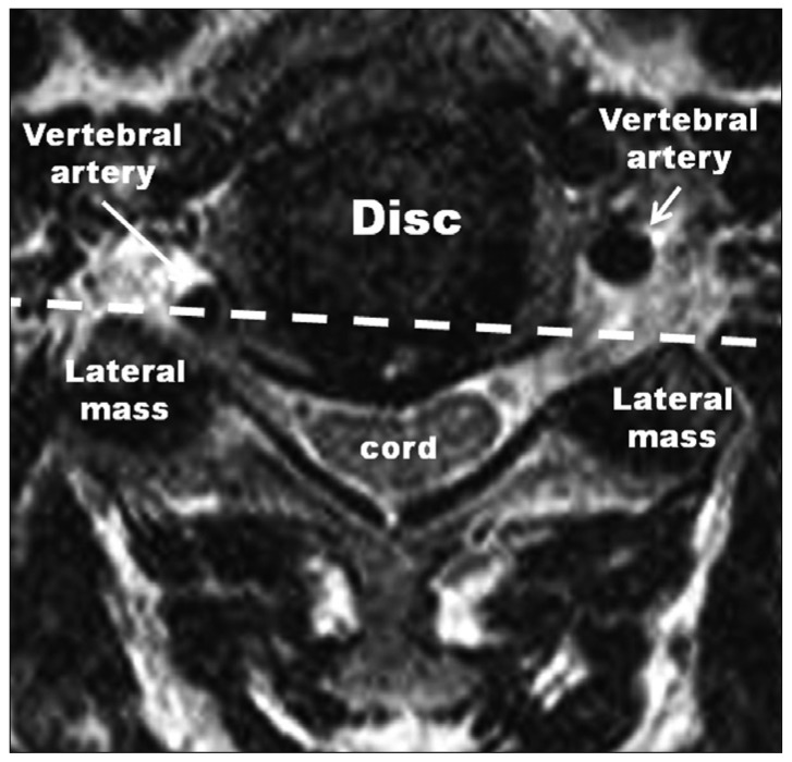
Fig. 1 This picture shows a transaxial section at the level of intervertebral disc. Over a third of the axial aspect of left vertebral artery located in the intervertebral foramen is inside the dotted line between most ventral points of bilateral lateral mass.
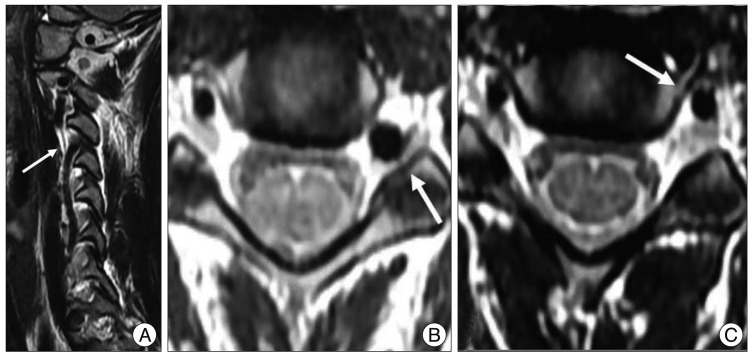
Fig. 2 Medial loop of vertebral artery on MRI scans. T2-weight sagittal MRI section shows a disappearance of vertebral artery at the level of the C3-4 foramen (white arrow) (A). T2-weight axial MRI scans show a abnormal position of the vertebral artery leading to compressing of the left C4 root at the level of the C3-4 foramen (white arrow) (B) and normal position of vertebral artery at the level of the C4-5 foramen (white arrow) (C).
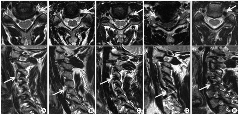
Fig. 3 The vertebral arteries form medial loops into intervertebral foramens and compress the proximal cervical roots (arrow). Upper and lower rows indicate the axial and sagittal section of T2 weight image in MRI scans. Fig. 3A to E corresponds with cases 2 to 6.
Reference
-
1. Aubin ME, Eskander MS, Drew JM, Marvin J, Eskander JP, Eck J, et al. Identification of type 1 : interforaminal vertebral artery anomalies in cervical spine MRIs. Spine (Phila Pa 1976). 2010; 35:E1610–E1611. PMID: 21116215.2. Bose B. Anterior cervical fusion using Caspar plating : analysis of results and review of the literature. Surg Neurol. 1998; 49:25–31. PMID: 9428891.3. Bruneau M, Cornelius JF, Marneffe V, Triffaux M, George B. Anatomical variations of the V2 segment of the vertebral artery. Neurosurgery. 2006; 59:ONS20–ONS24. discussion ONS20-ONS24. PMID: 16888547.
Article4. Chibbaro S, Mirone G, Yasuda M, Marsella M, Di Emidio P, George B. Vertebral artery loop--a cause of cervical radiculopathy. World Neurosurg. 2012; 78:375e11–e13. PMID: 22381311.
Article5. Curylo LJ, Mason HC, Bohlman HH, Yoo JU. Tortuous course of the vertebral artery and anterior cervical decompression : a cadaveric and clinical case study. Spine (Phila Pa 1976). 2000; 25:2860–2864. PMID: 11074670.
Article6. Ekinci G, Baltacioglu F, Ozgen S, Akpinar I, Erzen C, Pamir N. Cervical neural foraminal widening caused by the tortuous vertebral artery. Clin Imaging. 2001; 25:320–322. PMID: 11682288.
Article7. Garcia Alzamora M, Rosahl SK, Lehmberg J, Klisch J. Life-threatening bleeding from a vertebral artery pseudoaneurysm after anterior cervical spine approach : endovascular repair by a triple stent-in-stent method. Case report. Neuroradiology. 2005; 47:282–286. PMID: 15789201.
Article8. Haller JM, Iwanik M, Shen FH. Clinically relevant anatomy of recurrent laryngeal nerve. Spine (Phila Pa 1976). 2012; 37:97–100. PMID: 21540775.
Article9. Hong JT, Lee SW, Son BC, Sung JH, Yang SH, Kim IS, et al. Analysis of anatomical variations of bone and vascular structures around the posterior atlantal arch using three-dimensional computed tomography angiography. J Neurosurg Spine. 2008; 8:230–236. PMID: 18312074.
Article10. Hong JT, Park DK, Lee MJ, Kim SW, An HS. Anatomical variations of the vertebral artery segment in the lower cervical spine : analysis by three-dimensional computed tomography angiography. Spine (Phila Pa 1976). 2008; 33:2422–2426. PMID: 18923317.
Article11. Kim HS, Lee JH, Cheh G, Lee SH. Cervical radiculopathy caused by vertebral artery loop formation : a case report and review of the literature. J Korean Neurosurg Soc. 2010; 48:465–468. PMID: 21286489.
Article12. Matula C, Trattnig S, Tschabitscher M, Day JD, Koos WT. The course of the prevertebral segment of the vertebral artery : anatomy and clinical significance. Surg Neurol. 1997; 48:125–131. PMID: 9242236.13. Müller M, Bleeck J, Ruf M. Vertebral artery anomaly with entry at C4--avoiding a surgical pitfall : a case report. Eur Spine J. 2008; 17(Suppl 2):S291–S293. PMID: 18180962.14. Oga M, Yuge I, Terada K, Shimizu A, Sugioka Y. Tortuosity of the vertebral artery in patients with cervical spondylotic myelopathy. Risk factor for the vertebral artery injury during anterior cervical decompression. Spine (Phila Pa 1976). 1996; 21:1085–1089. PMID: 8724095.
Article15. Ozgen S, Pait TG, Cağlar YS. The V2 segment of the vertebral artery and its branches. J Neurosurg Spine. 2004; 1:299–305. PMID: 15478368.
Article16. Russo VM, Graziano F, Peris-Celda M, Russo A, Ulm AJ. The V(2) segment of the vertebral artery : anatomical considerations and surgical implications. J Neurosurg Spine. 2011; 15:610–619. PMID: 21905775.
Article17. Satti SR, Cerniglia CA, Koenigsberg RA. Cervical vertebral artery variations : an anatomic study. AJNR Am J Neuroradiol. 2007; 28:976–980. PMID: 17494682.
- Full Text Links
-
- Actions
-
Cited
- CITED
-
- Close
- Share
-
- Similar articles
-
- Cervical Radiculopathy Caused by Vertebral Artery Loop Formation : A Case Report and Review of the Literature
- Vertebral Artery Loop Formation Depicted on Oblique Sagittal MR Imaging: A Case Report
- Morphometric Measurement of the Anatomical Landmark in Anterior Cervical Microforaminotomy
- Emergent Endovascular Embolization for Iatrogenic Vertebral Artery Injury during Cervical Discectomy and Fusion
- A case of occipital artery originating from the vertebral artery
