Experimental Evaluation of Accelerated T1rho Relaxation Quantification in Human Liver Using Limited Spin-Lock Times
- Affiliations
-
- 1Department of Imaging and Interventional Radiology, The Chinese University of Hong Kong, Prince of Wales Hospital, Shatin, Hong Kong SAR, China. yixiang_wang@cuhk.edu.hk
- 2Department of Radiology, Zhongda Hospital, Southeast University, Nanjing 210009, China.
- KMID: 1397504
- DOI: http://doi.org/10.3348/kjr.2012.13.6.736
Abstract
OBJECTIVE
It was reported lately that to obtain consistent liver T1rho measurement, at 3T MRI using six spin-lock times (SLTs), is feasible. In this study, the feasibility of using three or two SLT points to measure liver T1rho relaxation time was explored.
MATERIALS AND METHODS
Seventeen healthy volunteers underwent 36 examinations. Three representative axial slices were selected to cut through the upper, middle, and lower liver. A rotary echo spin-lock pulse was implemented in a 2D fast field echo sequence. Spin-lock frequency was 500 Hz and the spin-lock times of 1, 10, 20, 30, 40, and 50 milliseconds (ms) were used for T1rho mapping. T1rho maps were constructed by using all 6 SLT points, three SLT points of 1, 20, and 50 ms, or two SLTs of 1 and 50 ms, respectively. Intra-class correlation coefficient (ICC) and Bland and Altman plot were used to assess the measurement agreement.
RESULTS
Two examinations were excluded, due to motion artifact at the SLT of 50 ms. With the remaining 34 examinations, the ICC for 6-SLT vs. 3-SLT T1rho measurements was 0.922, while the ICC for 6-SLT vs. 2-SLT T1rho measurement was 0.756. The Bland and Altman analysis showed a mean difference of 0.19 (95% limits of agreement: -1.34, 1.73) for 6-SLT vs. 3-SLT T1rho measurement, and the mean difference of 0.89 (95% limits of agreement: -1.67, 3.45) for 6-SLT vs. 2-SLT T1rho measurement. The scan re-scan reproducibility ICC (n = 11 subjects) was 0.755, 0.727, and 0.528 for 6-SLT measurement, 3-SLT measurement, and 2-SLT measurement, respectively.
CONCLUSION
Adopting 3 SLTs of 1, 20, and 50 ms can be an acceptable alternative for the liver T1rho measurement, while 2 SLTs of 1 and 50 ms do not provide reliable measurement.
Keyword
MeSH Terms
Figure
-
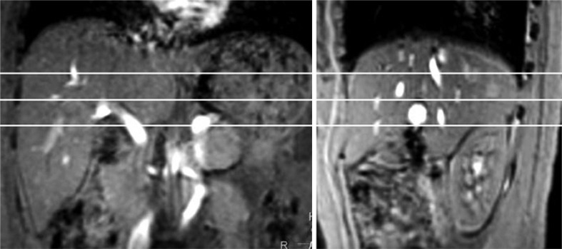
Fig. 1 Coronal (right) and sagittal (left) scout images. Three axial slices are selected to cut through upper, middle, and lower liver.
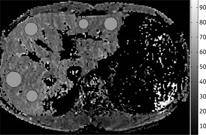
Fig. 2 T1rho map with coefficient of determination R2 > 0.8 evaluation. Five regions of interest are placed on liver parenchyma region, excluding observable artifacts and blood vessels.
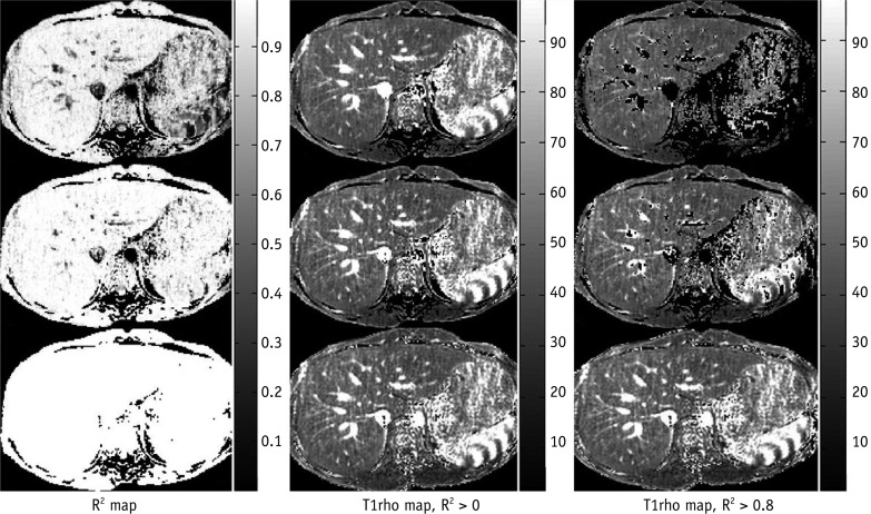
Fig. 3 Example of one healthy volunteer's T1rho maps constructed with 6 SLTs, 3 SLTs or 2 SLTs. Left column: coefficient of determination (R2) maps, middle column: T1rho maps without R2 evaluation, right column: T1rho maps with R2 > 0.8 evaluation. Upper row: maps constructed with 6 SLTs, middle row: maps constructed with 3 SLTs, lower row: maps constructed with 2 SLTs. As SLT point decreases, R2 evaluation is less efficient to remove blood vessel contamination and artifact. Note that R2 is always equal to one, when SLT point = 2, so no pixels are excluded by criteria of R2 > 0.8. However, this does not seem to affect regions of interest placement, according to results in Table 1. Radiology trainee was able to identify and avoid regions with artifacts and blood vessels. SLT = spin-lock time
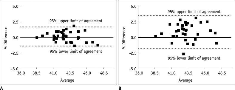
Fig. 4 Bland and Altman plots for comparison of 6 SLTs vs. 3 SLTs liver T1rho measurement (A) and 6 SLTs vs. 2 SLTs liver T1rho measurement (B). 3 SLTs measurement demonstrates good agreement with 6 SLTs measurement. SLT = spin-lock time
Cited by 1 articles
-
Impact of Liver Fibrosis and Fatty Liver on T1rho Measurements: A Prospective Study
Shuangshuang Xie, Qing Li, Yue Cheng, Yu Zhang, Zhizheng Zhuo, Guiming Zhao, Wen Shen
Korean J Radiol. 2017;18(6):898-905. doi: 10.3348/kjr.2017.18.6.898.
Reference
-
1. Afdhal NH, Nunes D. Evaluation of liver fibrosis: a concise review. Am J Gastroenterol. 2004; 99:1160–1174. PMID: 15180741.
Article2. Blumenkrantz G, Zuo J, Li X, Kornak J, Link TM, Majumdar S. In vivo 3.0-tesla magnetic resonance T1rho and T2 relaxation mapping in subjects with intervertebral disc degeneration and clinical symptoms. Magn Reson Med. 2010; 63:1193–1200. PMID: 20432290.3. Borthakur A, Sochor M, Davatzikos C, Trojanowski JQ, Clark CM. T1rho MRI of Alzheimer's disease. Neuroimage. 2008; 41:1199–1205. PMID: 18479942.4. Pakin SK, Xu J, Schweitzer ME, Regatte RR. Rapid 3D-T1rho mapping of the knee joint at 3.0T with parallel imaging. Magn Reson Med. 2006; 56:563–571. PMID: 16894582.5. Santyr GE, Henkelman RM, Bronskill MJ. Spin locking for magnetic resonance imaging with application to human breast. Magn Reson Med. 1989; 12:25–37. PMID: 2607958.
Article6. Virta A, Komu M, Lundbom N, Jääskeläinen S, Kalimo H, Airio A, et al. Low field T1rho imaging of myositis. Magn Reson Imaging. 1998; 16:385–391. PMID: 9665549.7. Lamminen AE, Tanttu JI, Sepponen RE, Pihko H, Korhola OA. T1 rho dispersion imaging of diseased muscle tissue. Br J Radiol. 1993; 66:783–787. PMID: 8220948.8. Wang YX, Yuan J, Chu ES, Go MY, Huang H, Ahuja AT, et al. T1rho MR imaging is sensitive to evaluate liver fibrosis: an experimental study in a rat biliary duct ligation model. Radiology. 2011; 259:712–719. PMID: 21436087.9. Sirlin CB. Science to practice: can T1rho imaging be used to diagnose and assess the severity of hepatic fibrosis? Radiology. 2011; 259:619–620. PMID: 21602499.10. Zhao F, Wang YX, Yuan J, Deng M, Wong HL, Chu ES, et al. MR T1ρ as an imaging biomarker for monitoring liver injury progression and regression: an experimental study in rats with carbon tetrachloride intoxication. Eur Radiol. 2012; 22:1709–1716. PMID: 22752522.
Article11. Deng M, Zhao F, Yuan J, Ahuja AT, Wang YX. Liver MR T1ρ measurement in healthy human subjects at 3 T: a preliminary study with a two-dimensional fast-field echo sequence. Br J Radiol. 2012; 85:e590–e595. PMID: 22422392.12. Charagundla SR, Borthakur A, Leigh JS, Reddy R. Artifacts in T(1rho)-weighted imaging: correction with a self-compensating spin-locking pulse. J Magn Reson. 2003; 162:113–121. PMID: 12762988.13. Li X, Han ET, Busse RF, Majumdar S. In vivo T(1rho) mapping in cartilage using 3D magnetization-prepared angle-modulated partitioned k-space spoiled gradient echo snapshots (3D MAPSS). Magn Reson Med. 2008; 59:298–307. PMID: 18228578.14. Fleiss JL. Reliability of measurement. The design and analysis of clinical experiments. 1986. New York: John Wiley & Sons.15. Santyr GE, Fairbanks EJ, Kelcz F, Sorenson JA. Off-resonance spin locking for MR imaging. Magn Reson Med. 1994; 32:43–51. PMID: 8084236.
Article16. Fleysher R, Fleysher L, Gonen O. The optimal MR acquisition strategy for exponential decay constants estimation. Magn Reson Imaging. 2008; 26:433–435. PMID: 18093779.
Article17. Cheon JE, Kim IO, Seo JK, Ko JS, Lee JM, Shin CI, et al. Clinical application of liver MR imaging in Wilson's disease. Korean J Radiol. 2010; 11:665–672. PMID: 21076593.
Article18. Faria SC, Ganesan K, Mwangi I, Shiehmorteza M, Viamonte B, Mazhar S, et al. MR imaging of liver fibrosis: current state of the art. Radiographics. 2009; 29:1615–1635. PMID: 19959511.
Article19. Shim JH, Yu JS, Chung JJ, Kim JH, Kim KW. Segmental difference of the hepatic fibrosis from chronic viral hepatitis due to hepatitis B versus C virus infection: comparison using dual contrast material-enhanced MRI. Korean J Radiol. 2011; 12:431–438. PMID: 21852903.
Article20. Talwalkar JA, Yin M, Fidler JL, Sanderson SO, Kamath PS, Ehman RL. Magnetic resonance imaging of hepatic fibrosis: emerging clinical applications. Hepatology. 2008; 47:332–342. PMID: 18161879.
Article
- Full Text Links
-
- Actions
-
Cited
- CITED
-
- Close
- Share
-
- Similar articles
-
- The Effects of Proteoglycan and Type II Collagen on T1rho Relaxation Time of Articular Cartilage
- Impact of Liver Fibrosis and Fatty Liver on T1rho Measurements: A Prospective Study
- T1-, T2-weighted, and FLAIR Imaging: Clinical Application
- Determination of Electron Spin Relaxation Time of the Gadolinium-Chealted MRI Contrast Agents by Using an X-band EPR Technique
- Imaging evaluation of non-alcoholic fatty liver disease: focused on quantification
