Revisit of Broden's View for Intraarticular Calcaneal Fracture
- Affiliations
-
- 1Department of Orthopaedic Surgery, Inha University Hospital, Incheon, Korea.
- 2Department of Orthopaedic Surgery, Seoul National University Bundang Hospital, Seongnam, Korea. oasis100@empal.com
- 3Department of Orthopaedic Surgery, Seoul National University Hospital, Seoul, Korea.
- KMID: 1392985
- DOI: http://doi.org/10.4055/cios.2012.4.3.221
Abstract
- BACKGROUND
This study was performed to investigate the relationship between coronal computed tomography (CT) and Broden's view in terms of location of the fracture line and fracture pattern.
METHODS
Forty-five feet of 45 patients with intraarticular calcaneal fractures were evaluated. The mean age of the patients was 46.3 years (standard deviation, 18.1; range, 15 to 80 years), and there were 34 men and 11 women. The Broden's views were acquired using the ray sum projection, reviewed, and correlated with the coronal CT image to determine the location of the fracture on the posterior facet and fracture pattern described by the Sanders classification. The quantified location of the fracture line was defined as the distance between the medial margin of posterior facet and the fracture line divided by the whole length of the posterior facet, which was expressed as a percentage.
RESULTS
The fracture line on the Broden's view was positioned at 22.3% (standard deviation, 29.6) laterally compared to that on coronal CT (p < 0.01). Although all cases showed posterior facet involvement on the CT scan, the fracture line was positioned lateral to the posterior facet in 6 cases (13.3%) in the Broden's view. The coronal CT and Broden's view showed a low level of agreement in the fracture pattern according to the Sanders classification, with kappa values of 0.23.
CONCLUSIONS
Surgeons should consider that the fracture line on the Broden's view shows positioning laterally compared to coronal CT and they should consider that the fracture line at the lateral to posterior facet on the Broden's view might be an intraarticular fracture line. There are some limitations when applying the Sanders classification with the Broden's view.
MeSH Terms
Figure
-
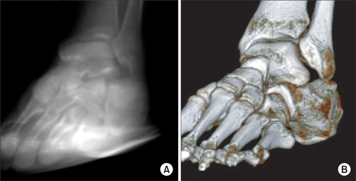
Fig. 1 Xelis ver. 1.0.2.2 enables the user to simulate different positions of the model, and gain the ray sum projection, which is most analogous in appearance to conventional radiographs from three-dimensional computed tomography (3D-CT) scans. This function was used to acquire the Broden's view. (A) After the ray sum projection to the foot and ankle model with an internal rotation of 30° was simulated, and an image similar to a conventional radiograph was gained. (B) The matched 3D-CT image is shown.
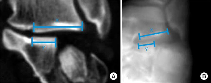
Fig. 2 The ratio of the total width of the posterior talocalcaneal articulation to the width between the first fracture line and the medial margin (Y/X) was measured on coronal computed tomography image (A) and Broden's view (B).
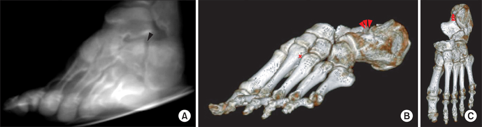
Fig. 3 Only posterior part of the sagittal fracture line is visible intraarticularly on the Broden's view. (A) Fracture line shown on reconstructed Broden's view image (black arrow head). (B) Three-dimensional computed tomography (3D-CT) image shows that the fracture line shown on the reconstructed Broden's view is the fracture gap in the summit of posterior facet, which is the posterior part of the fracture line (red arrow heads). Talus has been eliminated from the image using the Xelis ver. 1.0.2.2. (C) Top-down view of the same 3D image. Please note that the anterior part of the fracture line is not located as laterally as the posterior part of the fracture line (red arrow heads).
Reference
-
1. Gurtowski J, Ries MD, Levin PE. Dee R, Mango E, Hurst LC, editors. Fractures and dislocations of the foot. Principles of orthopaedic practice. 1989. New York: McGraw-Hill;1242–1260.2. Sangeorzan BJ, Benirschke SK, Carr JB. Surgical management of fractures of the os calcis. Instr Course Lect. 1995. 44:359–370.3. Eastwood DM, Gregg PJ, Atkins RM. Intra-articular fractures of the calcaneum. Part I: pathological anatomy and classification. J Bone Joint Surg Br. 1993. 75(2):183–188.
Article4. Sangeorzan BJ, Ananthakrishnan D, Tencer AF. Contact characteristics of the subtalar joint after a simulated calcaneus fracture. J Orthop Trauma. 1995. 9(3):251–258.
Article5. Thoren O. Os calcis fractures. Acta Orthop Scand Suppl. 1964. 70:Suppl 70. 1–116.6. Crosby LA, Kamins P. The history of the calcaneal fracture. Orthop Rev. 1991. 20(6):501–509.7. Buckley R, Tough S, McCormack R, et al. Operative compared with nonoperative treatment of displaced intra-articular calcaneal fractures: a prospective, randomized, controlled multicenter trial. J Bone Joint Surg Am. 2002. 84(10):1733–1744.
Article8. Janzen DL, Connell DG, Munk PL, Buckley RE, Meek RN, Schechter MT. Intraarticular fractures of the calcaneus: value of CT findings in determining prognosis. AJR Am J Roentgenol. 1992. 158(6):1271–1274.
Article9. Crosby LA, Fitzgibbons T. Computerized tomography scanning of acute intra-articular fractures of the calcaneus: a new classification system. J Bone Joint Surg Am. 1990. 72(6):852–859.
Article10. Ebraheim NA, Elgafy H, Sabry FF, Tao S. Calcaneus fractures with subluxation of the posterior facet: a surgical indication. Clin Orthop Relat Res. 2000. (377):210–216.11. Gilmer PW, Herzenberg J, Frank JL, Silverman P, Martinez S, Goldner JL. Computerized tomographic analysis of acute calcaneal fractures. Foot Ankle. 1986. 6(4):184–193.
Article12. Druckmann A, Schwartz A, Rabinovici N, Feuchtwanger M. Pneumatosis of the intestines. Am J Roentgenol Radium Ther Nucl Med. 1961. 86:911–919.13. Heger L, Wulff K. Computed tomography of the calcaneus: normal anatomy. AJR Am J Roentgenol. 1985. 145(1):123–129.
Article14. Heger L, Wulff K, Seddiqi MS. Computed tomography of calcaneal fractures. AJR Am J Roentgenol. 1985. 145(1):131–137.
Article15. Lowrie IG, Finlay DB, Brenkel IJ, Gregg PJ. Computerised tomographic assessment of the subtalar joint in calcaneal fractures. J Bone Joint Surg Br. 1988. 70(2):247–250.
Article16. Rosenberg ZS, Feldman F, Singson RD. Intra-articular calcaneal fractures: computed tomographic analysis. Skeletal Radiol. 1987. 16(2):105–113.
Article17. Sanders R. Intra-articular fractures of the calcaneus: present state of the art. J Orthop Trauma. 1992. 6(2):252–265.18. Sanders R. Displaced intra-articular fractures of the calcaneus. J Bone Joint Surg Am. 2000. 82(2):225–250.
Article19. Sanders R, Fortin P, DiPasquale T, Walling A. Operative treatment in 120 displaced intraarticular calcaneal fractures: results using a prognostic computed tomography scan classification. Clin Orthop Relat Res. 1993. (290):87–95.20. Segal D, Marsh JL, Leiter B. Clinical application of computerized axial tomography (CAT) scanning of calcaneus fractures. Clin Orthop Relat Res. 1985. (199):114–123.
Article21. Dalrymple NC, Prasad SR, Freckleton MW, Chintapalli KN. Informatics in radiology (infoRAD): introduction to the language of three-dimensional imaging with multidetector CT. Radiographics. 2005. 25(5):1409–1428.
Article22. Levine RD. Volume rendering with the Kubota 3D imaging and graphics accelerator. Digit Tech J. 1994. 6(2):34–48.23. Moshiri M, Scarfe WC, Hilgers ML, Scheetz JP, Silveira AM, Farman AG. Accuracy of linear measurements from imaging plate and lateral cephalometric images derived from cone-beam computed tomography. Am J Orthod Dentofacial Orthop. 2007. 132(4):550–560.
Article24. Reddinger WL. CT instrumentation & physics [Internet]. 1997. cited 2012 Jun 28. Longwood: OutSource Inc.;Available from: http://www.e-radiography.net/mrict/Basic_CT.pdf.25. Bonett DG. Sample size requirements for estimating intraclass correlations with desired precision. Stat Med. 2002. 21(9):1331–1335.
Article26. McGraw KO, Wong SP. Forming inferences about some intraclass correlations coefficients: correction. Psychol Methods. 1996. 1(4):390.
Article27. Landis JR, Koch GG. The measurement of observer agreement for categorical data. Biometrics. 1977. 33(1):159–174.
Article28. Allon SM. Fractures of the calcaneus. Foot Ankle Int. 1996. 17(11):720.29. Furey A, Stone C, Squire D, Harnett J. Os calcis fractures: analysis of interobserver variability in using Sanders classification. J Foot Ankle Surg. 2003. 42(1):21–23.
Article30. Lauder AJ, Inda DJ, Bott AM, Clare MP, Fitzgibbons TC, Mormino MA. Interobserver and intraobserver reliability of two classification systems for intra-articular calcaneal fractures. Foot Ankle Int. 2006. 27(4):251–255.
Article
- Full Text Links
-
- Actions
-
Cited
- CITED
-
- Close
- Share
-
- Similar articles
-
- The Role of Simple Radiography in the Evaluation of Intraarticular Calcaneal Fracture
- Minimally Invasive Reduction and Pin Fixation Treatment for Displaced Intraarticular Calcaneal Fracture
- Operative Treatment of Intraarticular Calcaneal Fracture: Comparison of Outcomes between Open Reduction and Closed Reduction
- Joint Depression Type of Intraarticular Calcaneal Fractures Treated with Essex-Lopresti Method
- Limited Posterior Approach for the Surgical Treatment of Intraarticular Fracture of Calcaneus
