Ventriculus Terminalis in Adults: Unusual Magnetic Resonance Imaging Features and Review of the Literature
- Affiliations
-
- 1Department of Radiology, Gangnam Severance Hospital, Yonsei University College of Medicine, Seoul 135-720, Korea. tschung@yuhs.ac
- 2Department of Radiology, Yonsei University College of Medicine, Seoul 120-752, Korea.
- 3Department of Neurosurgery, Gangnam Severance Hospital, Yonsei University College of Medicine, Seoul 135-720, Korea.
- KMID: 1392933
- DOI: http://doi.org/10.3348/kjr.2012.13.5.557
Abstract
OBJECTIVE
The ventriculus terminalis (VT) in adults is a rare pathology. We report various MR imaging features of the adult VT.
MATERIALS AND METHODS
Ten patients were included in this retrospective review.. All patients had undergone magnetic resonance (MR imaging with a surface coil that used two different 1.5T MR systems. All patients had undergone initial and follow-up MR imaging with contrast enhancement using gadopentate dimeglumine. Three patients underwent additional MR imaging using the echocardiogram-gated spatial modulation of magnetization (SPAMM) technique. If a shift in tagging band during the systolic phase was less than half of the band space, it was defined as a "non-pulsatile fluid". Two neuroradiologists independently reviewed these images, while clinical symptoms and outcomes were statistically analyzed between the treated and non-treated group.
RESULTS
All cases presented an intramedullary cystic lesion in the conus medullaris and showed the same signal intensity as CSF. Three VTs had intracystic septation and cord edema, which were pathologically confirmed after surgery; two of these were associated with kyphotic deformity and spinal arteriovenous malformation. SPAMM-MRI of 3 patients demonstrated non-pulsatile fluid motion within the VT. In the treated group, clinical symptoms improved better than the non-treated group.
CONCLUSION
The adult VT shows some unusual imaging features, including septation, cord edema, and coexistence of a spinal AVM, as well as the typical findings. Surgical maneuvers may be considered as a treatment option in adult VT with progressive neurological symptoms.
MeSH Terms
Figure
-
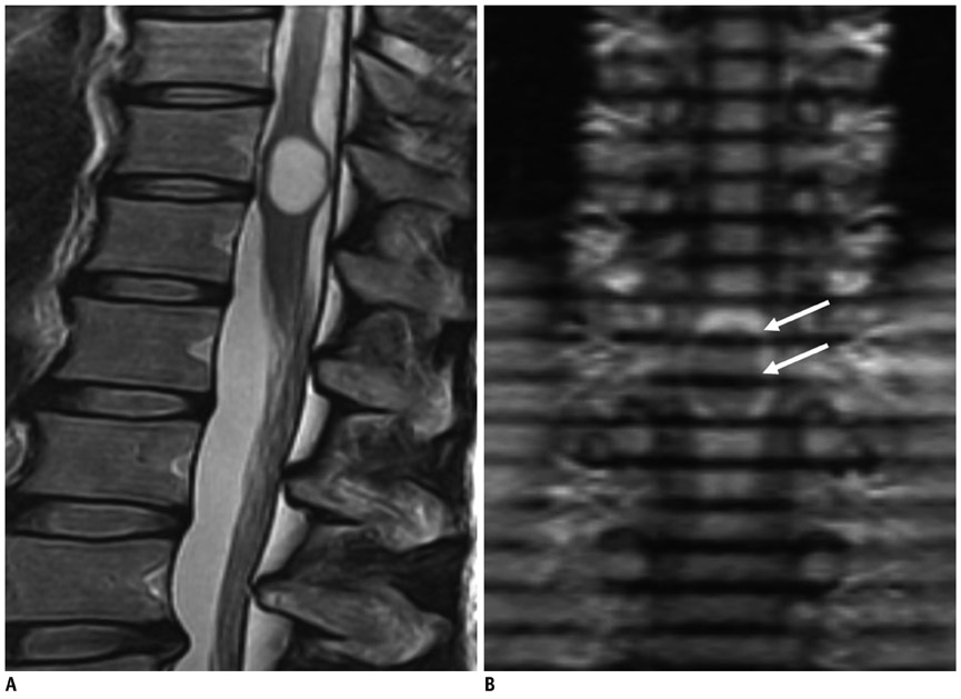
Fig. 1 43-year-old man presented with back pain. A. High signal-intensity T2-weighted sagittal image shows intramedullary cystic lesion in conus medullaris with. B. On spatial modulation of magnetization magnetic resonance sagittal image, there is no movement of tagging band in cystic ventriculus terminalis with cardiac pulsation (arrow).
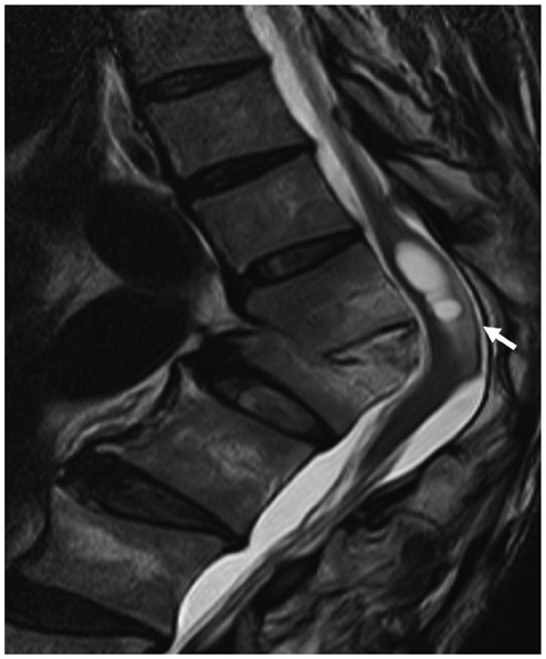
Fig. 2 50-year old man with left sciatic pain, who had kyphotic deformity. T2-weighted sagittal image shows cystic lesion of conus medullaris and accompanying cord edema (arrow) with thoracic kyphosis.
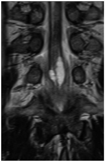
Fig. 3 T2-weighted coronal image shows cystic lesion of 33-year old man with urinary difficulty. Cystic lesion of conus medullaris has elements of septation and enlargement.
Cited by 1 articles
-
The terminal ventricle of Saguinus leucopus (Primate)
Jorge Eduardo Duque Parra, Miguel Alejandro Aguirre García, Juan Fernando Vélez García
Anat Cell Biol. 2020;53(4):502-504. doi: 10.5115/acb.20.062.
Reference
-
1. Unsinn KM, Mader R, Gassner I, Kreczy A, Freund MC. Sonography of the ventriculus terminalis in newborns. AJNR Am J Neuroradiol. 1996. 17:1003–1004.2. Kriss VM, Kriss TC, Babcock DS. The ventriculus terminalis of the spinal cord in the neonate: a normal variant on sonography. AJR Am J Roentgenol. 1995. 165:1491–1493.3. Coleman LT, Zimmerman RA, Rorke LB. Ventriculus terminalis of the conus medullaris: MR findings in children. AJNR Am J Neuroradiol. 1995. 16:1421–1426.4. de Moura Batista L, Acioly MA, Carvalho CH, Ebner FH, Tatagiba M. Cystic lesion of the ventriculus terminalis: proposal for a new clinical classification. J Neurosurg Spine. 2008. 8:163–168.5. Sigal R, Denys A, Halimi P, Shapeero L, Doyon D, Boudghène F. Ventriculus terminalis of the conus medullaris: MR imaging in four patients with congenital dilatation. AJNR Am J Neuroradiol. 1991. 12:733–737.6. Nassar SI, Correll JW, Housepian EM. Intramedullary cystic lesions of the conus medullaris. J Neurol Neurosurg Psychiatry. 1968. 31:106–109.7. Unsinn KM, Mader R, Gassner I, Kreczy A. Ventriculus terminalis of the spinal cord in the neonate: a normal variant on sonography. AJR Am J Roentgenol. 1996. 167:1341.8. Truong BC, Shaw DW, Winters WD. Dilation of the ventriculus terminalis: sonographic findings. J Ultrasound Med. 1998. 17:713–715.9. Kriss VM, Kriss TC, Coleman RC. Sonographic appearance of the ventriculus terminalis cyst in the neonatal spinal cord. J Ultrasound Med. 2000. 19:207–209.10. Kernohan JW. The ventriculus terminalis: its growth and development. J Comp Neurol. 1924. 38:107–125.11. Byrd SE, Harvey C, Darling CF. MR of terminal myelocystoceles. Eur J Radiol. 1995. 20:215–220.12. Taviere V, Brunelle F, Baraton J, Temam M, Pierre-Kahn A, Lallemand D. MRI study of lumbosacral lipoma in children. Pediatr Radiol. 1989. 19:316–320.13. Erkan K, Unal F, Kiris T. Terminal syringomyelia in association with the tethered cord syndrome. Neurosurgery. 1999. 45:1351–1359. discussion 1359-1360.14. Srivatanakul K, Songsaeng D, Ozanne A, Toulgoat F, Lasjaunias P. Spinal arteriovenous malformation associated with syringomyelia. J Neurosurg Spine. 2009. 10:436–442.15. Lee KH, Chung TS, Jeon TJ, Kim YH, Chien D, Laub G. Application of spatial modulation of magnetization to cervical spinal stenosis for evaluation of the hydrodynamic changes occurring in cerebrospinal fluid. Korean J Radiol. 2000. 1:11–18.16. Wayte SC, Redpath TW. Cine magnetic resonance imaging of pulsatile cerebrospinal fluid flow using CSPAMM. Br J Radiol. 1994. 67:1088–1095.17. Park CH, Chung TS, Kim DJ, Suh SH, Chung WS, Cho YE. Evaluation of intrasyrinx fluid motion by spatial modulation of magnetization-magnetic resonance imaging in syringomyelia with long-term follow-up: a predictor of postoperative prognosis? J Comput Assist Tomogr. 2008. 32:135–140.18. Sakas DE, Korfias SI, Wayte SC, Beale DJ, Papapetrou KP, Stranjalis GS, et al. Chiari malformation: CSF flow dynamics in the craniocervical junction and syrinx. Acta Neurochir (Wien). 2005. 147:1223–1233.19. Lee SK, Chung TS, Kim YS. Evaluation of CSF motion in syringomyelia with spatial modulation of magnetization (SPAMM). Yonsei Med J. 2002. 43:37–42.20. Seo HS, Kim JH, Lee DH, Lee YH, Suh SI, Kim SY, et al. Nonenhancing intramedullary astrocytomas and other MR imaging features: a retrospective study and systematic review. AJNR Am J Neuroradiol. 2010. 31:498–503.21. Brisman JL, Li M, Hamilton D, Mayberg MR, Newell DW. Cystic dilation of the conus ventriculus terminalis presenting as an acute cauda equina syndrome relieved by decompression and cyst drainage: case report. Neurosurgery. 2006. 58:E585. discussion E585.22. Dullerud R, Server A, Berg-Johnsen J. MR imaging of ventriculus terminalis of the conus medullaris. A report of two operated patients and a review of the literature. Acta Radiol. 2003. 44:444–446.23. Celli P, D'Andrea G, Trillò G, Roperto R, Acqui M, Ferrante L. Cyst of the medullary conus: malformative persistence of terminal ventricle or compressive dilatation? Neurosurg Rev. 2002. 25:103–106.24. Stewart DH Jr, King RB, Lourie H. Surgical drainage of cyst of the conus medullaris. Report of three cases. J Neurosurg. 1970. 33:106–110.25. Robertson DP, Kirkpatrick JB, Harper RL, Mawad ME. Spinal intramedullary ependymal cyst. Report of three cases. J Neurosurg. 1991. 75:312–316.26. Agrillo U, Tirendi MN, Nardi PV. Symptomatic cystic dilatation of V ventricle: case report and review of the literature. Eur Spine J. 1997. 6:281–283.27. Matsubayashi R, Uchino A, Kato A, Kudo S, Sakai S, Murata S. Cystic dilatation of ventriculus terminalis in adults: MRI. Neuroradiology. 1998. 40:45–47.28. Liccardo G, Ruggeri F, De Cerchio L, Floris R, Lunardi P. Fifth ventricle: an unusual cystic lesion of the conus medullaris. Spinal Cord. 2005. 43:381–384.29. Ciappetta P, D'urso PI, Luzzi S, Ingravallo G, Cimmino A, Resta L. Cystic dilation of the ventriculus terminalis in adults. J Neurosurg Spine. 2008. 8:92–99.
- Full Text Links
-
- Actions
-
Cited
- CITED
-
- Close
- Share
-
- Similar articles
-
- Developmental Changes of Glial Fibrillary Acidic Protein (GFAP) and Proliferating Cell Nuclear Antigen (PCNA) Immunoreactivity of the Ependyma lining the Central Canal and Ventriculus Terminalis in the Human Fetus
- Intramedullary Spinal Lesions Involving the Conus Medullaris: MR Imaging Features for Differential Diagnosis
- Morphological Study of the Ventriculus Terminalis in the Human Fetal Spinal Cord
- Bone Involvement of Diffuse Large B Cell Lymphoma (DLBCL) Showing Unusual Manifestations Mimicking Chronic Osteomyelitis in a 58-Year-Old Man: Case Report and Clinical Application of Diffusion Weighted Magnetic Resonance Imaging
- US or MR Imaging Features of Polypoid Endometriosis: A Case Report
