Korean J Radiol.
2012 Oct;13(5):550-556. 10.3348/kjr.2012.13.5.550.
Artificial Luminal Narrowing on Contrast-Enhanced Magnetic Resonance Angiograms on an Occasion of Stent-Assisted Coiling of Intracranial Aneurysm: In Vitro Comparison Using Two Different Stents with Variable Imaging Parameters
- Affiliations
-
- 1Department of Radiology, Seoul St. Mary's Hospital, The Catholic University of Korea College of Medicine, Seoul 137-701, Korea. bumrad@catholic.ac.kr
- 2Department of Neurosurgery, Seoul St. Mary's Hospital, The Catholic University of Korea College of Medicine, Seoul 137-701, Korea.
- KMID: 1392932
- DOI: http://doi.org/10.3348/kjr.2012.13.5.550
Abstract
OBJECTIVE
Intracranial stenting for stent-assisted coiling of aneurysms requires adequate follow-up imaging. The aim of this in vitro study was to compare in-stent artificial luminal narrowing on contrast-enhanced MR angiograms (CE-MRA) when applying Neuroform(R) and Enterprise(R) stents for stent-assisted coiling.
MATERIALS AND METHODS
Two intracranial nitinol stents (Enterprise(R) and Neuroform(R)) were placed in silicon tubes and then imaged at 3 T and 1.5 T by the use of a T1-weighted three-dimensional spoiled gradient-echo sequence with minimal TR and TE. CE-MRAs were obtained by using different imaging planes, voxel sizes, and bandwidths, and with or without parallel imaging. Artificial lumen narrowing (ALN) was calculated and the results were compared.
RESULTS
Lower magnetic field strength, axial plane perpendicular to axis of stent, and wider bandwidth resulted in a lower ALN on CE-MRA for both stents. Larger voxel size resulted in lower ALN for Neuroform(R) stent. The parallel imaging acceleration factor did not affect ALN. The mean ALN was lower for Neuroform(R), but it was not significant by a paired t test.
CONCLUSION
CE-MRA of the stented lumen of vascular phantom was partially impaired with ALN. Consequently, image plane orientation, magnetic field strength, bandwidth, and voxel size should be adjusted appropriately to reduce ALN.
MeSH Terms
Figure
-
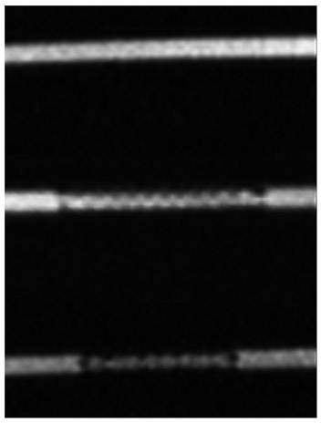
Fig. 1 Static artifact appearance on series 3 (coronal orientation; 3T; TR/TE, 4.2/1.6; voxel size, 0.7 × 0.7 × 0.7 mm3; slice thickness, 0.7 mm; bandwidth, 430 Hz/pixel; FOV, 113 × 300; matrix size, 168 × 448; no parallel imaging acceleration factor). Images were obtained as control tube, Enterprise® and Neuroform® from top to bottom. TR = repetition time, TE = echo time
Reference
-
1. Bartels LW, Bakker CJ, Viergever MA. Improved lumen visualization in metallic vascular implants by reducing RF artifacts. Magn Reson Med. 2002. 47:171–180.2. Fabregues S, Baijens K, Rieu R, Bergeron P. Hemodynamics of endovascular prostheses. J Biomech. 1998. 31:45–54.3. Müller-Hülsbeck S, Schwarzenberg H, Wesner F, Drost R, Glüer CC, Heller M. Visualization of flow patterns from stents and stent-grafts in an in vitro flow-model. Invest Radiol. 1998. 33:762–770.4. Choi JW, Roh HG, Moon WJ, Chun YI, Kang CH. Optimization of MR Parameters of 3D TOF-MRA for Various Intracranial Stents at 3.0T MRI. Neurointervention. 2011. 6:71–77.5. Huang BY, Castillo M. Neurovascular imaging at 1.5 tesla versus 3.0 tesla. Magn Reson Imaging Clin N Am. 2009. 17:29–46.6. Blum MB, Schmook M, Schernthaner R, Edelhauser G, Puchner S, Lammer J, et al. Quantification and detectability of in-stent stenosis with CT angiography and MR angiography in arterial stents in vitro. AJR Am J Roentgenol. 2007. 189:1238–1242.7. Borisch I, Hamer OW, Zorger N, Feuerbach S, Link J. In vivo evaluation of the carotid wallstent on three-dimensional contrast material-enhanced MR angiography: influence of artifacts on the visibility of stent lumina. J Vasc Interv Radiol. 2005. 16:669–677.8. Hagspiel KD, Leung DA, Nandalur KR, Angle JF, Dulai HS, Spinosa DJ, et al. Contrast-enhanced MR angiography at 1.5 T after implantation of platinum stents: in vitro and in vivo comparison with conventional stent designs. AJR Am J Roentgenol. 2005. 184:288–294.9. Hähnel S, Nguyen-Trong TH, Rohde S, Hartmann M, Braun C, Sartor K, et al. 3.0 Tesla contrast-enhanced MR angiography of carotid artery stents: in vitro measurements as compared with 1.5 Tesla. J Neuroradiol. 2006. 33:75–80.10. Hamer OW, Borisch I, Paetzel C, Nitz WR, Seitz J, Feuerbach S, et al. In vitro evaluation of stent patency and in-stent stenoses in 10 metallic stents using MR angiography. Br J Radiol. 2006. 79:636–643.11. Klemm T, Duda S, Machann J, Seekamp-Rahn K, Schnieder L, Claussen CD, et al. MR imaging in the presence of vascular stents: A systematic assessment of artifacts for various stent orientations, sequence types, and field strengths. J Magn Reson Imaging. 2000. 12:606–615.12. Maintz D, Kugel H, Schellhammer F, Landwehr P. In vitro evaluation of intravascular stent artifacts in three-dimensional MR angiography. Invest Radiol. 2001. 36:218–224.13. Straube T, Wolf S, Flesser A, Deli M, Alfke K, Nabavi A, et al. [MRI of carotid stents: influence of stent properties and sequence parameters on visualization of the carotid artery lumen]. Rofo. 2005. 177:375–380.14. Wall A, Kugel H, Bachman R, Matuszewski L, Krämer S, Heindel W, et al. 3.0 T vs. 1.5 T MR angiography: in vitro comparison of intravascular stent artifacts. J Magn Reson Imaging. 2005. 22:772–779.15. Frölich AM, Pilgram-Pastor SM, Psychogios MN, Mohr A, Knauth M. Comparing different MR angiography strategies of carotid stents in a vascular flow model: toward stent-specific recommendations in MR follow-up. Neuroradiology. 2011. 53:359–365.16. Lettau M, Sauer A, Heiland S, Rohde S, Bendszus M, Hähnel S. Carotid artery stents: in vitro comparison of different stent designs and sizes using CT angiography and contrast-enhanced MR angiography at 1.5T and 3T. AJNR Am J Neuroradiol. 2009. 30:1993–1997.17. Lettau M, Sauer A, Heiland S, Rohde S, Reinhardt J, Bendszus M, et al. In vitro comparison of different carotid artery stents: a pixel-by-pixel analysis using CT angiography and contrast-enhanced MR angiography at 1.5 and 3 T. Neuroradiology. 2010. 52:823–830.18. Bartels LW, Smits HF, Bakker CJ, Viergever MA. MR imaging of vascular stents: effects of susceptibility, flow, and radiofrequency eddy currents. J Vasc Interv Radiol. 2001. 12:365–371.19. Meyer JM, Buecker A, Spuentrup E, Schuermann K, Huetten M, Hilgers RD, et al. Improved in-stent magnetic resonance angiography with high flip angle excitation. Invest Radiol. 2001. 36:677–681.20. Wajnberg E, de Souza JM, Marchiori E, Gasparetto EL. Single-center experience with the Neuroform stent for endovascular treatment of wide-necked intracranial aneurysms. Surg Neurol. 2009. 72:612–619.21. Peluso JP, van Rooij WJ, Sluzewski M, Beute GN. A new self-expandable nitinol stent for the treatment of wide-neck aneurysms: initial clinical experience. AJNR Am J Neuroradiol. 2008. 29:1405–1408.22. Biondi A, Janardhan V, Katz JM, Salvaggio K, Riina HA, Gobin YP. Neuroform stent-assisted coil embolization of wide-neck intracranial aneurysms: strategies in stent deployment and midterm follow-up. Neurosurgery. 2007. 61:460–468. discussion 468-469.23. Lee YJ, Kim DJ, Suh SH, Lee SK, Kim J, Kim DI. Stent-assisted coil embolization of intracranial wide-necked aneurysms. Neuroradiology. 2005. 47:680–689.24. Schenck JF. The role of magnetic susceptibility in magnetic resonance imaging: MRI magnetic compatibility of the first and second kinds. Med Phys. 1996. 23:815–850.25. Bakker CJ, Bhagwandien R, Moerland MA, Fuderer M. Susceptibility artifacts in 2DFT spin-echo and gradient-echo imaging: the cylinder model revisited. Magn Reson Imaging. 1993. 11:539–548.26. Lüdeke KM, Röschmann P, Tischler R. Susceptibility artefacts in NMR imaging. Magn Reson Imaging. 1985. 3:329–343.27. Camacho CR, Plewes DB, Henkelman RM. Nonsusceptibility artifacts due to metallic objects in MR imaging. J Magn Reson Imaging. 1995. 5:75–88.28. Lenhart M, Völk M, Manke C, Nitz WR, Strotzer M, Feuerbach S, et al. Stent appearance at contrast-enhanced MR angiography: in vitro examination with 14 stents. Radiology. 2000. 217:173–178.
- Full Text Links
-
- Actions
-
Cited
- CITED
-
- Close
- Share
-
- Similar articles
-
- Delayed Self-expansion Phenomenon as a Complication of Neuroform Stent Assisted Coiling for Ruptured Intracranial Aneurysm
- PulseRider Treated Aneurysm with Significant Artifact on Postoperative Magnetic Resonance Angiography: A Case Report and Literature Review
- A Complicated Case of Endovascular Stent Assisted Coil Embolization of an Aneurysm
- Very Late Stent Thrombosis after Sole Stent-Assisted Coiling at the Paraclinoid Giant Aneurysm : Could Prophylactic Antiplatelet Therapy Be Ceased at the Only 1 Year after Procedure?
- Y-Stenting Endovascular Treatment for Ruptured Intracranial Aneurysms : A Single-Institution Experience in Korea
