J Vet Sci.
2012 Sep;13(3):327-330. 10.4142/jvs.2012.13.3.327.
Use of canine small intestinal submucosa allograft for treating perineal hernias in two dogs
- Affiliations
-
- 1Department of Veterinary Surgery, College of Veterinary Medicine, Konkuk University, Seoul 143-701, Korea. hykim@konkuk.ac.kr
- 2Department of Veterinary Clinical Pathology, College of Veterinary Medicine, Konkuk University, Seoul 143-701, Korea.
- 3Department of Veterinary Radiology, College of Veterinary Medicine, Konkuk University, Seoul 143-701, Korea.
- KMID: 1389774
- DOI: http://doi.org/10.4142/jvs.2012.13.3.327
Abstract
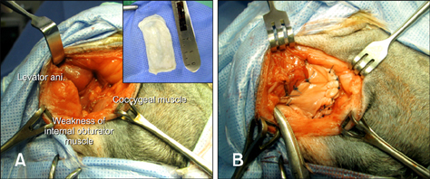
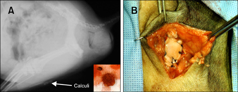
- Here, we describe two dogs in which canine small intestinal submucosa (SIS) was implanted as a biomaterial scaffold during perineal herniorrhaphy. Both dogs had developed severe muscle weakness, unilaterally herniated rectal protrusions, and heart problems with potential anesthetic risks. Areas affected by the perineal hernia (PH) located between the internal obturator and external anal sphincter muscles were reconstructed with naive canine SIS sheets. In 12 months, post-operative complications such as wound infections, sciatic paralysis, rectal prolapse, or recurrence of the hernia were not observed. Symptoms of defecatory tenesmus also improved. Neither case showed any signs of rejection or specific immune responses as determined by complete and differential cell counts. Our findings demonstrate that canine SIS can be used as a biomaterial scaffold for PH repair in dogs.
MeSH Terms
Figure
Reference
-
1. Anderson MA, Constantinescu GM, Mann FA. Bojrab MJ, Ellison GW, Slocum B, editors. Perineal hernia repair in the dog. Current Techniques in Small Animal Surgery. 1998. 4th ed. Philadelphia: Lippincott Williams & Wilkins;555–564.2. Badylak SF, Lantz GC, Coffey A, Geddes LA. Small intestinal submucosa as a large diameter vascular graft in the dog. J Surg Res. 1989. 47:74–80.
Article3. Bellenger CR. Perineal hernia in dogs. Aust Vet J. 1980. 56:434–438.
Article4. Bellenger CR, Canfield RB. Slatter DH, editor. Perineal hernia. Textbook of Small Animal Surgery. 2003. 3rd ed. Philadelphia: Saunders;487–498.5. Burrows CF, Harvey CE. Perineal hernia in the dog. J Small Anim Pract. 1973. 14:315–332.
Article6. Clarke KM, Lantz GC, Salisbury SK, Badylak SF, Hiles MC, Voytik SL. Intestine submucosa and polypropylene mesh for abdominal wall repair in dogs. J Surg Res. 1996. 60:107–114.
Article7. Dorn AS, Cartee RE, Richardson DC. A preliminary comparison of perineal hernia in the dog and man. J Am Anim Hosp Assoc. 1982. 18:624–632.8. Frankland AL. Use of porcine dermal collagen in the repair of perineal hernia in dogs-a preliminary report. Vet Rec. 1986. 119:13–14.
Article9. Hardie EM, Kolata RJ, Earley TD, Rawlings CA, Gorgacz EJ. Evaluation of internal obturator muscle transposition in treatment of perineal hernia in dogs. Vet Surg. 1983. 12:69–72.
Article10. Hodde JP, Badylak SF, Brightman AO, Voytik-Harbin SL. Glycosaminoglycan content of small intestinal submucosa: a bioscaffold for tissue replacement. Tissue Eng. 1996. 2:209–217.
Article11. Kropp BP, Eppley BL, Prevel CD, Rippy MK, Harruff RC, Badylak SF, Adams MC, Rink RC, Keating MA. Experimental assessment of small intestinal submucosa as a bladder wall substitute. Urology. 1995. 46:396–400.
Article12. Matthiesen DT. Diagnosis and management of complications occurring after perineal herniorrhaphy in dogs. Compend Contin Educ Pract Vet. 1989. 11:797–802.13. Niebauer GW, Shibly S, Seltenhammer M, Pirker A, Brandt S. Relaxin of prostatic origin might be linked to perineal hernia formation in dogs. Ann N Y Acad Sci. 2005. 1041:415–422.
Article14. Novitsky YW, Harrell AG, Cristiano JA, Paton BL, Norton HJ, Peindl RD, Kercher KW, Heniford BT. Comparative evaluation of adhesion formation, strength of ingrowth, and textile properties of prosthetic meshes after long-term intra-abdominal implantation in a rabbit. J Surg Res. 2007. 140:6–11.
Article15. Raffan PJ. A new surgical technique for repair of perineal hernias in the dog. J Small Anim Pract. 1993. 34:13–19.
Article16. Schumpelick V, Klinge U. Prosthetic implants for hernia repair. Br J Surg. 2003. 90:1457–1458.
Article17. Shahar R, Shamir MH, Niebauer GW, Johnston DE. A possible association between acquired nontraumatic inguinal and perineal hernia in adult male dogs. Can Vet J. 1996. 37:614–616.18. Sjollema BE, van Sluijs FJ. Perineal hernia repair in the dog by transposition of the internal obturator muscle. II. Complications and results in 100 patients. Vet Q. 1989. 11:18–23.
Article19. Stoll MR, Cook JL, Pope ER, Carson WL, Kreeger JM. The use of porcine small intestinal submucosa as a biomaterial for perineal herniorrhaphy in the dog. Vet Surg. 2002. 31:379–390.
Article20. Voytik-Harbin SL, Brightman AO, Kraine MR, Waisner B, Badylak SF. Identification of extractable growth factors from small intestinal submucosa. J Cell Biochem. 1997. 67:478–491.
Article
- Full Text Links
- Actions
-
Cited
- CITED
-
- Close
- Share
- Similar articles
-
- Evaluation of a canine small intestinal submucosal xenograft and polypropylene mesh as bioscaffolds in an abdominal full-thickness resection model of growing rats
- Application of pulsed Doppler ultrasound for the evaluation of small intestinal motility in dogs
- Spontaneous Transomental Hernia
- Silicone stenting as an emergency option for treatment of canine laryngeal paralysis
- Prevalence of Giardia intestinalis and other zoonotic intestinal parasites in private household dogs of the Hachinohe area in Aomori prefecture, Japan in 1997, 2002 and 2007


