J Vet Sci.
2012 Sep;13(3):311-314. 10.4142/jvs.2012.13.3.311.
Contrast-enhanced ultrasound analysis of renal perfusion in normal micropigs
- Affiliations
-
- 1Department of Veterinary Medical Imaging, and Research Institute for Veterinary Science, College of Veterinary Medicine, Seoul National University, Seoul 151-742, Korea. mcchoi@snu.ac.kr
- KMID: 1389771
- DOI: http://doi.org/10.4142/jvs.2012.13.3.311
Abstract
- Contrast-enhanced ultrasound is one of method for evaluating renal perfusion. The purpose of this project was to assess perfusion patterns and dynamics in normal micropig kidney using ultrasonographic contrast media. Eight young healthy micropigs were included in this study. Micropigs were anesthetized with propofol and received an intravenous bolus of microbubble contrast media through an ear vein. Time/mean pixel value (MPV) curves were generated for selected regions in the right renal cortex and medulla. The parenchyma was enhanced in two phases. The cortex was first enhanced followed by a more gradual enhancement of the medulla. A significant difference in perfusion was detected between the cortex and medulla. Following the bolus injection, the average upslope was 0.68 +/- 0.27 MPV/sec, downslope was -0.27 +/- 0.13 MPV/sec, baseline was 73.9 +/- 16.5 MPV, peak was 84.6 +/- 17.2 MPV, and time-to-peak (from injection) was 17.5 +/- 6.6 sec for the cortex. For the medulla, the average upslope was 0.50 +/- 0.24 MPV/sec, downslope was -0.12 +/- 0.06 MPV/sec, baseline was 52.7 +/- 7.0 MPV, peak was 65.2 +/- 9.3 MPV, and time-to-peak (from injection) was 27.5 +/- 5.0 sec. These data can be used as normal reference values for studying young micropigs.
Keyword
MeSH Terms
-
Animals
Contrast Media/*diagnostic use
Image Processing, Computer-Assisted
Injections, Intravenous/veterinary
Kidney/*blood supply/ultrasonography
Kidney Function Tests/veterinary
Linear Models
Microbubbles/diagnostic use/veterinary
Reference Values
Renal Circulation
Sulfur Hexafluoride/diagnostic use
Swine
Swine, Miniature/*physiology
Ultrasonography/*methods/veterinary
Figure
-
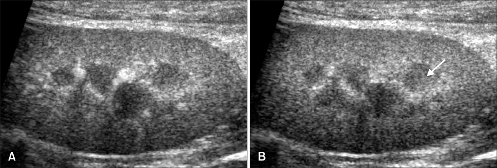
Fig. 1 (A) Representative longitudinal plane images of the renal cortex captured at the time of peak intensity using contrast-enhanced ultrasonography. Maximum contrast enhancement of the renal cortex occurred at 17.5 ± 6.6 sec. (B) The renal medulla was also enhanced (arrow) and had a uniformly hyperechoic appearance.
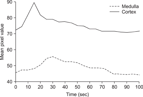
Fig. 2 Representative time-mean pixel value (MPV) curves obtained by contrast-enhanced ultrasonography in the renal cortex and medulla of a healthy micropig. The upper curve represents enhancement of the cortex and the lower curve shows enhancement of the medulla.
Reference
-
1. Ascenti G, Zimbaro G, Mazziotti S, Chimenz R, Fede C, Visalli C, Scribano E. Harmonic US imaging of vesicoureteric reflux in children: usefulness of a second generation US contrast agent. Pediatr Radiol. 2004. 34:481–487.
Article2. Bouakaz A, de Jong N. WFUMB Safety symposium on echo-contrast agents: nature and types of ultrasound contrast agents. Ultrasound Med Biol. 2007. 33:187–196.
Article3. Claudon M, Barnewolt CE, Taylor GA, Dunning PS, Gobet R, Badawy AB. Renal blood flow in pigs: changes depicted with contrast-enhanced harmonic US imaging during acute urinary obstruction. Radiology. 1999. 212:725–731.
Article4. Evans HE, Christensen GC. Evans HE, editor. The urogenital system. Miller's Anatomy of the Dog. 1993. 3rd ed. Philadelphia: WB Saunders;494–503.5. Hollenberg N. Black D, Jones NF, editors. The physiology of the renal circulation. Renal Disease. 1979. 4th ed. Oxford: Blackwell;30–63.6. Jayaweera AR, Edwards N, Glasheen WP, Villanueva FS, Abbott RD, Kaul S. In vivo myocardial kinetics of air-filled albumin microbubbles during myocardial contrast echocardiography. Comparison with radiolabeled red blood cells. Circ Res. 1994. 74:1157–1165.
Article7. Keller MW, Segal SS, Kaul S, Duling B. The behavior of sonicated albumin microbubbles within the microcirculation: a basis for their use during myocardial contrast echocardiography. Circ Res. 1989. 65:458–467.
Article8. Kim B, Lim HK, Choi MH, Woo JY, Ryu J, Kim S, Peck KR. Detection of parenchymal abnormalities in acute pyelonephritis by pulse inversion harmonic imaging with or without microbubble ultrasonographic contrast agent. Correlation with computed tomography. J Ultrasound Med. 2001. 20:5–14.
Article9. Kinns J, Aronson L, Hauptman J, Seiler G. Contrast-enhanced ultrasound of the feline kidney. Vet Radiol Ultrasound. 2010. 51:168–172.
Article10. Leinonen MR, Raekallio MR, Vainio OM, Ruohoniemi MO, O'Brien RT. The effect of the sample size and location on contrast ultrasound measurement of perfusion parameters. Vet Radiol Ultrasound. 2011. 52:82–87.
Article11. Miller DL, Dou C, Wiggins RC. Contrast-enhanced diagnostic ultrasound causes renal tissue damage in a porcine model. J Ultrasound Med. 2010. 29:1391–1401.
Article12. Nyman HT, Kristensen AT, Kjelgaard-Hansen M, McEvoy FJ. Contrast-enhanced ultrasonography in normal canine liver. Evaluation of imaging and safety parameters. Vet Radiol Ultrasound. 2005. 46:243–250.
Article13. O'Brien RT, Iani M, Matheson J, Delaney F, Young K. Contrast harmonic ultrasound of spontaneous liver nodules in 32 dogs. Vet Radiol Ultrasound. 2004. 45:547–553.14. Pallone TL, Zhang Z, Rhinehart K. Physiology of the renal medullary microcirculation. Am J Physiol Renal Physiol. 2003. 284:F253–F266.
Article15. Pietra M, Brini E, Fracassi F, Diana A, Cipone M. Use of the galactose-based contrast agent SHU 508A (Levovist) in renal ultrasonography of the dog. Vet Res Commun. 2005. 29:Suppl 2. 305–307.
Article16. Quaia E. Microbubble ultrasound contrast agents: an update. Eur Radiol. 2007. 17:1995–2008.
Article17. Rademacher N, Ohlerth S, Scharf G, Laluhova D, Sieber-Ruckstuhl N, Alt M, Roos M, Grest P, Kaser-Hotz B. Contrast-enhanced power and color Doppler ultrasonography of the pancreas in healthy and diseased cats. J Vet Intern Med. 2008. 22:1310–1316.
Article18. Waller KR, O'Brien RT, Zagzebski JA. Quantitative contrast ultrasound analysis of renal perfusion in normal dogs. Vet Radiol Ultrasound. 2007. 48:373–377.
Article19. Wei K, Le E, Bin JP, Coggins M, Thorpe J, Kaul S. Quantification of renal blood flow with contrast-enhanced ultrasound. J Am Coll Cardiol. 2001. 37:1135–1140.
Article
- Full Text Links
-
- Actions
-
Cited
- CITED
-
- Close
- Share
-
- Similar articles
-
- Renal Perfusion Image Using Harmonic Ultrasound with Microbble Contrast Agent: Preliminary Study
- Contrast-enhanced ultrasound of hepatocellular carcinoma: where do we stand?
- The Study of Renal Perfusion Image in Rabbit by Harmonic Ultrasound with Microbble Contrast Agent in Comparison with 99mTc-DTPA: Focusing on US Scan Technique and Concentration of Contrast Agent
- Contrast Enhanced Ultrasound Perfusion Imaging in Skeletal Muscle
- A Primer on the Methods and Applications for Contrast Echocardiography in Clinical Imaging
