Korean J Ophthalmol.
2012 Jun;26(3):230-234. 10.3341/kjo.2012.26.3.230.
Indocyanine Green Angiographic Findings of Obscure Choroidal Abnormalities in Neurofibromatosis
- Affiliations
-
- 1Department of Ophthalmology and Visual Science, Seoul St. Mary's Hospital, The Catholic University of Korea College of Medicine, Seoul, Korea. nuntangi5@gmail.com
- KMID: 1376132
- DOI: http://doi.org/10.3341/kjo.2012.26.3.230
Abstract
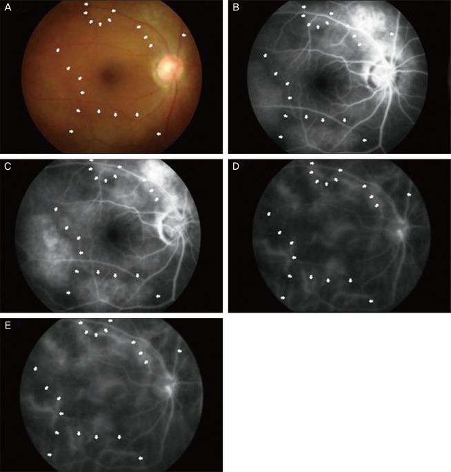
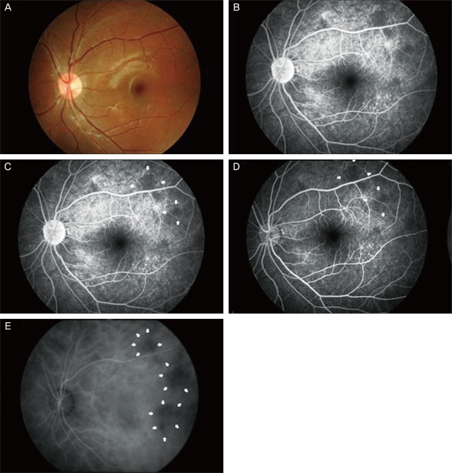
- We report two cases of choroidal neurofibromatosis, detected with the aid of indocyanine green angiography (ICGA) in patients with neurofibromatosis (NF)-1, otherwise having obscure findings based on ophthalmoscopy and fluoresceine angiography (FA). In case 1, the ophthalmoscopic exam showed diffuse bright or yellowish patched areas with irregular and blunt borders at the posterior pole. The FA showed multiple hyperfluorescent areas at the posterior pole in the early phase, which then showed more hyperfluorescence without leakage or extent in the late phase. The ICGA showed diffuse hypofluorescent areas in both the early and late phases, and the deep choroidal vessels were also visible. In case 2, the fundus showed no abnormal findings, and the FA showed weakly hypofluorescent areas with indefinite borders in both eyes. With the ICGA, these areas were more hypofluorescent and had clear borders. Choroidal involvement in NF-1 seems to occur more than expected. In selected cases, ICGA is a useful tool to be utilized when an ocular examination is conducted in a patient that has no definite findings based on the ophthalmoscope, B-scan, or FA tests.
MeSH Terms
Figure
Reference
-
1. Von Recklinghausen FD. Ueber die multiplen Fibrome der Haut und ihre Beziehung zu den multiplen Neuromen. Festschrift zur Feier des fünfundzwanzigjährigen Bestehens des pathologischen Instituts zu Berlin. Herrn Rudolf Virchow. 1882. Berlin: Hirschwald.2. National Institutes of Health Consensus Development Conference. Neurofibromatosis: conference statement. Arch Neurol. 1988; 45:575–578. PMID: 3128965.3. Martuza RL, Eldridge R. Neurofibromatosis 2 (bilateral acoustic neurofibromatosis). N Engl J Med. 1988; 318:684–688. PMID: 3125435.4. DeBella K, Szudek J, Friedman JM. Use of the national institutes of health criteria for diagnosis of neurofibromatosis 1 in children. Pediatrics. 2000; 105(3 Pt 1):608–614. PMID: 10699117.
Article5. Lewis RA, Riccardi VM. Von Recklinghausen neurofibromatosis: incidence of iris hamartomata. Ophthalmology. 1981; 88:348–354. PMID: 6789269.6. Destro M, D'Amico DJ, Gragoudas ES, et al. Retinal manifestations of neurofibromatosis. Diagnosis and management. Arch Ophthalmol. 1991; 109:662–666. PMID: 1902661.7. Martyn LJ, Knox DL. Glial hamartoma of the retina in generalized neurofibromatosis, Von Recklinghausen's disease. Br J Ophthalmol. 1972; 56:487–491. PMID: 4627012.
Article8. Palmer ML, Carney MD, Combs JL. Combined hamartomas of the retinal pigment epithelium and retina. Retina. 1990; 10:33–36. PMID: 2343189.
Article9. Klein RM, Glassman L. Neurofibromatosis of the choroid. Am J Ophthalmol. 1985; 99:367–368. PMID: 3919589.
Article10. Rescaldani C, Nicolini P, Fatigati G, Bottoni FG. Clinical application of digital indocyanine green angiography in choroidal neurofibromatosis. Ophthalmologica. 1998; 212:99–104. PMID: 9486548.
Article11. Woog JJ, Albert DM, Craft J, et al. Choroidal ganglioneuroma in neurofibromatosis. Graefes Arch Clin Exp Ophthalmol. 1983; 220:25–31. PMID: 6403410.
Article12. Kurosawa A, Kurosawa H. Ovoid bodies in choroidal neurofibromatosis. Arch Ophthalmol. 1982; 100:1939–1941. PMID: 6816197.
Article13. Wolter JR. Nerve fibrils in ovoid bodies. Arch Ophthalmol. 1965; 73:696–699. PMID: 14281990.
Article14. Ferry AP. Ryan SJ, editor. Other phakomatoses. Retina. Vol. 1. Basic science and inherited retinal disease. 1994. 2nd ed. St Louis: Mosby;p. 650–659.15. Wolter JR, Gonzales-Sirit R, Mankin WJ. Neuro-fibromatosis of the choroid. Am J Ophthalmol. 1962; 54:217–225. PMID: 14040313.16. Albert DM. Garner A, Klintworth GK, editors. Tumors: nature and biology. Pathobiology of ocular disease. 1982. Vol. 1. New York: N. Dekker;p. 689–704.17. Flower RW, Hochheimer BF. A clinical technique and apparatus for simultaneous angiography of the separate retinal and choroidal circulations. Invest Ophthalmol. 1973; 12:248–261. PMID: 4632821.18. Bischoff PM, Flower RW. Ten years experience with choroidal angiography using indocyanine green dye: a new routine examination or an epilogue? Doc Ophthalmol. 1985; 60:235–291. PMID: 2414083.
Article19. Hayashi K, de Laey JJ. Indocyanine green angiography of submacular choroidal vessels in the human eye. Ophthalmologica. 1985; 190:20–29. PMID: 3969259.
Article20. Destro M, Puliafito CA. Indocyanine green videoangiography of choroidal neovascularization. Ophthalmology. 1989; 96:846–853. PMID: 2472588.
Article
- Full Text Links
- Actions
-
Cited
- CITED
-
- Close
- Share
- Similar articles
-
- Indocyanine Green Angiographics Findings in Central Serous Chorioretinopathy
- Indocyanine Green Angiographic Findings in Harada Disease
- Fluorescein and Indocyanine Green Angiography of Choroidal Tumors
- Availability of Optical Coherence Tomography in Diagnosis and Classification of Choroidal Neovascularization
- Angiographic Findings of Retinal Pigment Epithelial Tear in Age-related Macular Degeneration


