Korean J Urol.
2008 Nov;49(11):1018-1023. 10.4111/kju.2008.49.11.1018.
Appropriate Follow-up Ultrasonography Interval after Pyeloplasty in Children with Ureteropelvic Junction Obstruction
- Affiliations
-
- 1Department of Urology, Urological Science Institute, Yonsei University College of Medicine, Seoul, Korea. swhan@yuhs.ac
- 2Department of Urology, Hankook General Hospital, Cheongju, Korea.
- KMID: 1328175
- DOI: http://doi.org/10.4111/kju.2008.49.11.1018
Abstract
-
PURPOSE: Serial ultrasonography(US) is routinely performed after pyeloplasty in the setting of pediatric ureteropelvic junction obstruction(UPJO). We evaluated the adequacy of the follow-up US interval we are currently using, which calls for US at 1, 3, 6, 9, and 12 months following surgery.
MATERIALS AND METHODS
Between January 2002 and August 2005, 102 patients underwent dismembered pyeloplasty for unilateral UPJO. Within this group, we selected 95 patients with high grade hydronephrosis to participate in this study. The degree of hydronephrosis was graded according to the classification issued by the Society for Fetal Urology(SFU). Improvement was defined as at least one grade of reduction. Serial sonograms were performed at 1, 3, 6, 9, and 12 months postoperatively.
RESULTS
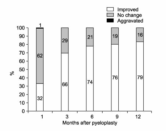
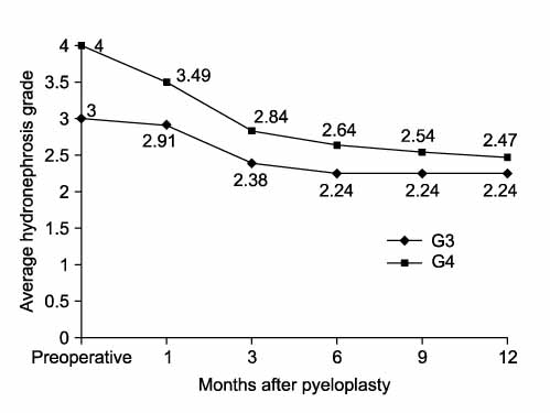
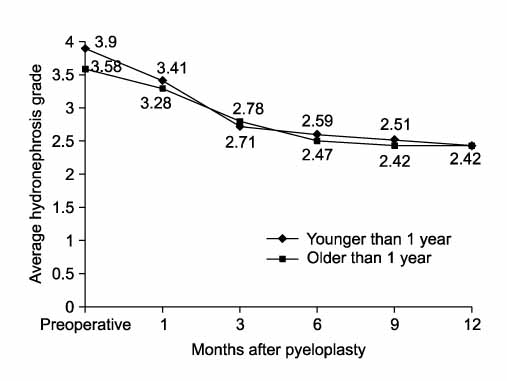
On follow-up US, 33.7%, 69.5%, 77.9%, 80.0%, and 83.2% of the patients showed improvement in their hydronephrosis at 1, 3, 6, 9, and 12 months, respectively. One patient presented with aggravation at 1 month. However, at 3 months, this patient had returned to the preoperative grade. There was no significant difference between the mean hydronephrosis grades at 6 and 12 months. No patient showed hydronephrosis aggravation at the 12-month follow-up examination.
CONCLUSIONS
US at 1, 3, and 6 months revealed significant improvements in hydronephrosis. However, no significant change in hydronephrosis occurred beyond 6 months. Therefore, US performed between 6 and 12 months after pyeloplasty may be inefficient, and we propose follow-up US at the following time points: 1 month, 3 to 6 months, 12 months, and then annually.
Figure
Reference
-
1. Fernbach SK, Maizels M, Conway JJ. Ultrasound grading of hydronephrosis: introduction to the system used by the Society for Fetal Urology. Pediatr Radiol. 1993. 23:478–480.2. Carr MC, El-ghoneimi A. Wein AJ, Kavoussi LR, Novick AC, Partin AW, Peters CA, editors. Anomalies and surgery of the ureteropelvic junction in children. Campbell-Walsh urology. 2007. 9th ed. Philadelphia: Saunders;3359–3382.3. Amling CL, O'Hara SM, Wiener JS, Schaeffer CS, King LR. Renal ultrasound changes after pyeloplasty in children with ureteropelvic junction obstruction: long-term outcome in 47 renal units. J Urol. 1996. 156:2020–2024.4. Dowling KJ, Harmon EP, Ortenberg J, Polanco E, Evans BB. Ureteropelvic junction obstruction: the effect of pyeloplasty on renal function. J Urol. 1988. 140:1227–1230.5. O'Reilly PH. Functional outcome of pyeloplasty for ureteropelvic junction obstruction: prospective study in 30 consecutive cases. J Urol. 1989. 142:273–276.6. Neste MG, du Cret RP, Finlay DE, Sane S, Gonzalez R, Boudreau RJ, et al. Postoperative diuresis renography and ultrasound in patients undergoing pyeloplasty. Predictors of surgical outcome. Clin Nucl Med. 1993. 18:872–876.7. Tapia J, Gonzalez R. Pyeloplasty improves renal function and somatic growth in children with ureteropelvic junction obstruction. J Urol. 1995. 154:218–222.8. Houben CH, Wischermann A, Börner G, Slany E. Outcome analysis of pyeloplasty in infants. Pediatr Surg Int. 2000. 16:189–193.9. Ulman I, Jayanthi VR, Koff SA. The long-term followup of newborns with severe unilateral hydronephrosis initially treated nonoperatively. J Urol. 2000. 164:1101–1105.10. Egan SC, Stock JA, Hanna MK. Renal ultrasound changes after internal double-J stented pyeloplasty for ureteropelvic junction obstruction. Tech Urol. 2001. 7:276–280.11. Konda R, Sakai K, Ota S, Abe Y, Hatakeyama T, Orikasa S. Ultrasound grade of hydronephrosis and severity of renal cortical damage on 99m technetium dimercaptosuccinic acid renal scan in infants with unilateral hydronephrosis during followup and after pyeloplasty. J Urol. 2002. 167:2159–2163.12. Palmer LS, Maizels M, Cartwright PC, Fernbach SK, Conway JJ. Surgery versus observation for managing obstructive grade 3 to 4 unilateral hydronephrosis: a report from the Society for Fetal Urology. J Urol. 1998. 159:222–228.13. Kim JW, Han SW, Choi SK. The postoperative prognosis of ureteropelvic junction obstruction according to the appearance of the ureter of preoperative retrograde pyelography. Korean J Urol. 2003. 44:550–555.14. Sheu JC, Koh CC, Chang PY, Wang NL, Tsai JD, Tsai TC. Ureteropelvic junction obstruction in children: 10 years' experience in one institution. Pediatr Surg Int. 2006. 22:519–523.15. Park S, Ji YH, Park YS, Kim KS. Change of hydronephrosis after pyeloplasty in children with unilateral ureteropelvic junction obstruction. Korean J Urol. 2005. 46:586–592.
- Full Text Links
- Actions
-
Cited
- CITED
-
- Close
- Share
- Similar articles
-
- Dismembered Pyeloplasty for Ureteropelvic Junction Obstruction in Children
- Change of Hydronephrosis after Pyeloplasty in Children with Unilateral Ureteropelvic Junction Obstruction
- Experiences on Anderson-Hynes Pyeloplasty
- Management of the Ureteropelvic Junction Obstruction
- Pyeloplasty for ureteropelvic junction obstruction : To divert or not to divert


