Unusual Thymic Hyperplasia Mimicking Lipomatous Tumor in an Eight-Year-Old Boy with Concomitant Pericardial Lipomatosis and Right Facial Hemihypertrophy
- Affiliations
-
- 1Department of Radiology, Seoul National University College of Medicine and the Institute of Radiation Medicine, Seoul 110-744, Korea. kimws@radcom.snu.ac.kr
- 2Department of Pathology, Seoul National University Hospital, Seoul National University College of Medicine, Seoul 110-744, Korea.
- KMID: 1122335
- DOI: http://doi.org/10.3348/kjr.2011.12.3.376
Abstract
- We report a case of thymic hyperplasia accompanied by pericardial lipomatosis and right facial hemihypertrophy in an 8-year-old boy. On imaging studies, the hyperplastic thymus had prominent curvilinear and nodular fatty areas simulating a fat-containing anterior mediastinal mass, which is an unusual finding in children. To our knowledge, this is the first report on a child with a combination of thymic hyperplasia, pericardial lipomatosis, and right facial hemihypertrophy. The radiologic findings are presented with a brief discussion.
Keyword
MeSH Terms
-
Child
Diagnosis, Differential
Facial Asymmetry/complications/*diagnosis
Heart Diseases/complications/*diagnosis/surgery
Humans
Hypertrophy/pathology
Lipomatosis/complications/*diagnosis/surgery
Magnetic Resonance Imaging
Male
Pericardium/*pathology
Thymus Hyperplasia/complications/*diagnosis/surgery
Tomography, X-Ray Computed
Figure
-
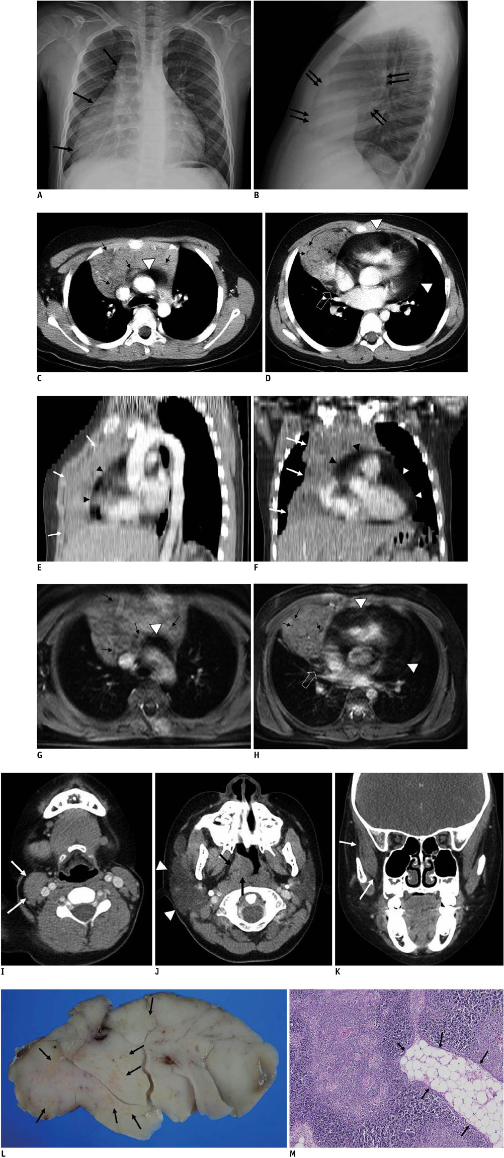
Fig. 1 8-year-old boy who had right facial hemihypertrophy, thymic hyperplasia and pericardial lipomatosis. A. Chest posteroanterior film shows mass-like shadow (black arrows) draping around and obliterating right cardiac border extending towards costophrenic angle. B. On chest lateral image, mass-like shadow is filling anterior mediastinal space (double arrows). C, D. On contrast-enhanced chest CT, axial image reveals large thymic mass showing predominant enlargement of right lobe with no definite mass effect on adjacent structures. Mass shows heterogeneous iso-attenuation to that of chest wall muscles containing areas of curvilinear (black arrows) and nodular (open arrows) fat density foci which was also more prominent on right lobe (C). Diffuse homogeneous soft tissue lesion with fat attenuation is filling pericardial space (arrowheads), suggestive of pericardial lipomatosis (D). E, F. Predominant enlargement of right thymic lobe (white arrows) and pericardial lipomatosis (arrowheads) is also well demonstrated on reconstructed oblique sagittal (E) and coronal images (F) of contrast-enhanced chest CT. G, H. Mediastinal MRI with contrast-enhanced fat-saturated T1-weighted image shows suppression of curvilinear (black arrows) or nodular (open arrows) areas within thymic mass and pericardial space (arrowheads), suggestive of areas of fatty component. I-K. On contrast-enhanced paranasal sinus (PNS) CT, thickening of right sternocleidomastoid muscle (arrows) (I) is noted on axial image. Right parotid gland (arrowheads) and right adenoid (arrows) is also enlarged (J). Coronal image reveals thickening of right temporalis muscle (arrows) (K). L. On gross specimen, cut surface of thymic mass is soft and yellow with no evidence of cystic change, necrosis or hemorrhage. Light-yellow spots (arrows) of multifocal fatty tissue are scattered within mass. M. On microscopic examination, mass shows histologic composition of normal thymus composed of cortical and medullary components containing islands of fatty tissue (arrows) (Hematoxylin & Eosin staining × 100).
Reference
-
1. Thomas LS. The mediastinum, Caffey's pediatric diagnostic imaging. 2007. 11th ed. Philadelphia: Mosby Elsevier;1339–1344.2. Baron RL, Lee JK, Sagel SS, Peterson RR. Computed tomography of the normal thymus. Radiology. 1982. 142:121–125.3. Siegel MJ, Glazer HS, Wiener JI, Molina PL. Normal and abnormal thymus in childhood: MR imaging. Radiology. 1989. 172:367–371.4. Takahashi K, Inaoka T, Murakami N, Hirota H, Iwata K, Nagasawa K, et al. Characterization of the normal and hyperplastic thymus on chemical-shift MR imaging. AJR Am J Roentgenol. 2003. 180:1265–1269.5. Nishino M, Ashiku SK, Kocher ON, Thurer RL, Boiselle PM, Hatabu H. The thymus: a comprehensive review. Radiographics. 2006. 26:335–348.6. Akhtar A, D'Cruz IA, Ramanathan KB, Umpierrez M. Diffuse locally invasive lipomatosis of the pericardium. Echocardiography. 2003. 20:173–177.7. Sanal HT, Kocaoglu M, Yildirim D, Ors F. Multiple cardiac lipomas and pericardial lipomatosis: multidetector-row computer tomography findings. Int J Cardiovasc Imaging. 2007. 23:655–658.8. Thiffault I, Schwartz CE, Der Kaloustian V, Foulkes WD. Mutation analysis of the tumor suppressor PTEN and the glypican 3 (GPC3) gene in patients diagnosed with Proteus syndrome. Am J Med Genet A. 2004. 130A:123–127.9. Dalal AB, Phadke SR, Pradhan M, Sharda S. Hemihyperplasia syndromes. Indian J Pediatr. 2006. 73:609–615.10. Boybeyi O, Alanay Y, Kayikcioglu A, Karnak I. Hemihyperplasia-multiple lipomatosis syndrome: an underdiagnosed entity in children with asymmetric overgrowth. J Pediatr Surg. 2010. 45:E19–E23.
