Effects of Propranolol on the Left Ventricular Volume of Normal Subjects During CT Coronary Angiography
- Affiliations
-
- 1Institute of Biomedical Engineering, College of Engineering and the College of Medicine, National Taiwan University, Taipei, Taiwan.
- 2Department of Radiology, Cathay General Hospital, Taipei, Taiwan.
- 3College of Medicine, Fu Jen Catholic University, Taipei, Taiwan.
- 4Department of Medical Imaging, National Taiwan University Hospital, Taipei, Taiwan. jamesfkp@yahoo.com.tw
- KMID: 1122329
- DOI: http://doi.org/10.3348/kjr.2011.12.3.319
Abstract
OBJECTIVE
The purpose of this study is to determine the effects of propranolol on the left ventricular (LV) volume during CT coronary angiography.
MATERIALS AND METHODS
The LV volume of 252 normal Chinese subjects (126 subjects with propranolol medication and 126 age- and gender-matched Chinese subjects without medication) was estimated using 64 slices multi-detector CT (MDCT). The heart rate difference was analyzed by the logistic linear regression model with variables that included gender, age, body height, body weight, systolic blood pressure (SBP), diastolic blood pressure (DBP) and the dosage of propranolol. The following global LV functional parameters were calculated: the real-end diastolic volume (EDV), the real-end systolic volume (ESV) and the real-ejection fraction (EF).
RESULTS
The female subjects had a greater decrease of heart rate after taking propranolol. The difference of heart rate was negatively correlated with the dosage of propranolol. The real-EDV, the real-ESV and the real-EF ranged from 48.1 to 109 mL/m2, 6.1 to 57.1 mL/m2 and 41% to 88%, respectively. There was no significant difference in the SBP and DBP between the groups without and with propranolol medication (123 +/- 17 and 80 +/- 10 mmHg; 120 +/- 14 and 80 +/- 11 mmHg, respectively). The real-EDV showed no significant difference between these two groups, but the real-ESV and real-EF showed significant differences between these two groups (69.4 +/- 9.3 and 70.6 +/- 8.9 mL/m2; 23.5 +/- 5.7 and 25.6 +/- 3.7 mL/m2, 66.5 +/- 5.1% and 63.5 +/- 4.6%, respectively).
CONCLUSION
The difference of heart rate is significantly influenced by gender and the dosage of propranolol. Propranolol will also increase the ESV, which contributes to a decreased EF, while the SBP, DBP and EDV are not statistically changed.
Keyword
MeSH Terms
-
Adrenergic beta-Antagonists/*administration & dosage
Case-Control Studies
China
Contrast Media/diagnostic use
*Coronary Angiography
Diastole
Electrocardiography
Female
Heart Rate/*drug effects
Humans
Logistic Models
Male
Middle Aged
Propranolol/*administration & dosage
Radiographic Image Interpretation, Computer-Assisted
Systole
*Tomography, X-Ray Computed
Triiodobenzoic Acids/diagnostic use
Ventricular Function, Left/*drug effects
Figure
-
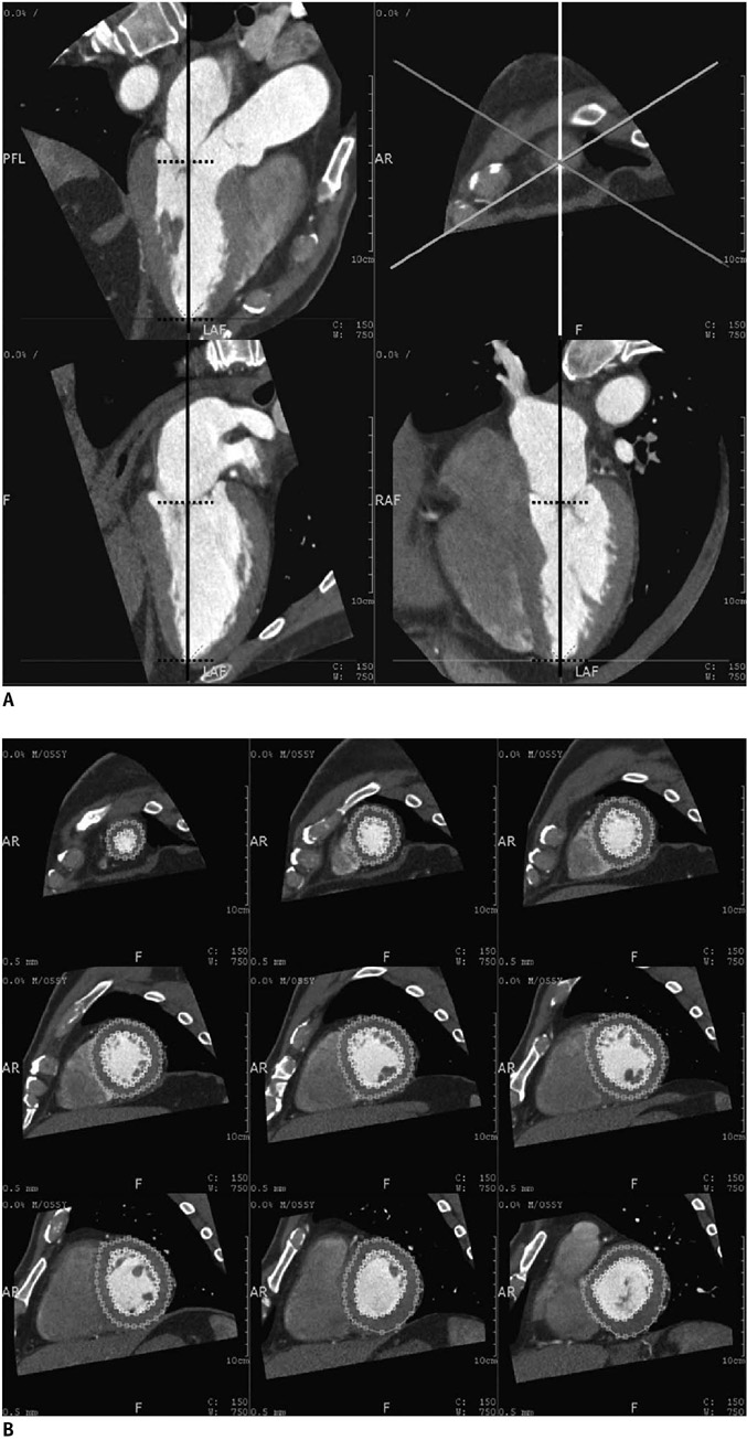
Fig. 1 Left ventricular volume of twenty phases' estimation. A. Cardiac axial images were automatically calculated and displayed on workstation. If necessary, we corrected axis by rotating vertical long axis (black solid line) to make axis orthogonal to plane of mitral valve and it passed center of mitral valve and apex. We defined area of interest by designating cardiac apex and concave center of mitral valve on planar images (dash horizontal short lines). B. Nine-slices of short-axis sections and contours of endocardial border of left ventricle were automatically traced and when necessary, traced contours were edited manually. Volume of papillary muscles was included in volume of ventricle.
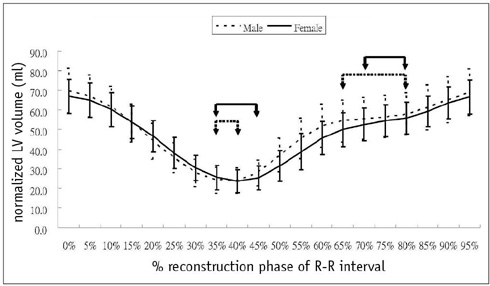
Fig. 2 Mean volume-time curve obtained from data of twenty phases' reconstruction. 35-40% and 65-80% phases of R-R interval in males (dash line) and 35-45% and 70-80% phases in females (solid line) showed smallest slope, which indicated least volumetric change.
Cited by 3 articles
-
Time Efficiency and Diagnostic Accuracy of New Automated Myocardial Perfusion Analysis Software in 320-Row CT Cardiac Imaging
Matthias Rief, Fabian Stenzel, Anisha Kranz, Peter Schlattmann, Marc Dewey
Korean J Radiol. 2013;14(1):21-29. doi: 10.3348/kjr.2013.14.1.21.Left Ventricular Functional Parameters and Geometric Patterns in Korean Adults on Coronary CT Angiography with a 320-Detector-Row CT Scanner
Eun-Ju Kang, Ki-Nam Lee, Won Jin Choi, Young-Dae Kim, Kyung Min Shin, Jae-Kwang Lim, Jongmin Lee
Korean J Radiol. 2017;18(4):664-673. doi: 10.3348/kjr.2017.18.4.664.Estimation of Diastolic Filling Pressure with Cardiac CT in Comparison with Echocardiography Using Tissue Doppler Imaging: Determination of Optimal CT Reconstruction Parameters
Ji-Sun Hwang, Heon Lee, Bora Lee, Soo-Jeong Lee, Sung Shick Jou, Hyun Kyung Lim, Jon Suh
Korean J Radiol. 2017;18(4):632-642. doi: 10.3348/kjr.2017.18.4.632.
Reference
-
1. Leschka S, Alkadhi H, Plass A, Desbiolles L, Grunenfelder J, Marincek B, et al. Accuracy of MSCT coronary angiography with 64-slice technology: first experience. Eur Heart J. 2005. 26:1482–1487.2. Raff GL, Gallagher MJ, O'Neill WW, Goldstein JA. Diagnostic accuracy of noninvasive coronary angiography using 64-slice spiral computed tomography. J Am Coll Cardiol. 2005. 46:552–557.3. Mollet NR, Cademartiri F, van Mieghem CA, Runza G, McFadden EP, Baks T, et al. High-resolution spiral computed tomography coronary angiography in patients referred for diagnostic conventional coronary angiography. Circulation. 2005. 112:2318–2323.4. Nikolaou K, Knez A, Rist C, Wintersperger BJ, Leber A, Johnson T, et al. Accuracy of 64-MDCT in the diagnosis of ischemic heart disease. AJR Am J Roentgenol. 2006. 187:111–117.5. Budoff MJ, Achenbach S, Blumenthal RS, Carr JJ, Goldin JG, Greenland P, et al. Assessment of coronary artery disease by cardiac computed tomography: a scientific statement from the American Heart Association Committee on Cardiovascular Imaging and Intervention, Council on Cardiovascular Radiology and Intervention, and Committee on Cardiac Imaging, Council on Clinical Cardiology. Circulation. 2006. 114:1761–1791.6. Stolzmann P, Scheffel H, Trindade PT, Plass AR, Husmann L, Leschka S, et al. Left ventricular and left atrial dimensions and volumes: comparison between dual-source CT and echocardiography. Invest Radiol. 2008. 43:284–289.7. van der Vleuten PA, Willems TP, Gotte MJ, Tio RA, Greuter MJ, Zijlstra F, et al. Quantification of global left ventricular function: comparison of multidetector computed tomography and magnetic resonance imaging. a meta-analysis and review of the current literature. Acta Radiol. 2006. 47:1049–1057.8. Wu YW, Tadamura E, Yamamuro M, Kanao S, Okayama S, Ozasa N, et al. Estimation of global and regional cardiac function using 64-slice computed tomography: a comparison study with echocardiography, gated-SPECT and cardiovascular magnetic resonance. Int J Cardiol. 2008. 128:69–76.9. Puesken M, Fischbach R, Wenker M, Seifarth H, Maintz D, Heindel W, et al. Global left-ventricular function assessment using dual-source multidetector CT: effect of improved temporal resolution on ventricular volume measurement. Eur Radiol. 2008. 18:2087–2094.10. Busch S, Johnson TR, Wintersperger BJ, Minaifar N, Bhargava A, Rist C, et al. Quantitative assessment of left ventricular function with dual-source CT in comparison to cardiac magnetic resonance imaging: initial findings. Eur Radiol. 2008. 18:570–575.11. Bastarrika G, Arraiza M, De Cecco CN, Mastrobuoni S, Ubilla M, Rabago G. Quantification of left ventricular function and mass in heart transplant recipients using dual-source CT and MRI: initial clinical experience. Eur Radiol. 2008. 18:1784–1790.12. Bansal D, Singh RM, Sarkar M, Sureddi R, McBreen KC, Griffis T, et al. Assessment of left ventricular function: comparison of cardiac multidetector-row computed tomography with two-dimension standard echocardiography for assessment of left ventricular function. Int J Cardiovasc Imaging. 2008. 24:317–325.13. Schlosser T, Mohrs OK, Magedanz A, Voigtlander T, Schmermund A, Barkhausen J. Assessment of left ventricular function and mass in patients undergoing computed tomography (CT) coronary angiography using 64-detector-row CT: comparison to magnetic resonance imaging. Acta Radiol. 2007. 48:30–35.14. Nakamura K, Funabashi N, Uehara M, Suzuki K, Terao M, Okubo K, et al. Quantitative 4-dimensional volumetric analysis of left ventricle in ischemic heart disease by 64-slice computed tomography: a comparative study with invasive left ventriculogram. Int J Cardiol. 2008. 129:42–52.15. Chaosuwannakit N, Rerkpattanapipat P, Wangsuphachart S, Srimahachota S. Reliability of the evaluation for left ventricular ejection fraction by ECG-gated multi-detector CT (MDCT): comparison with biplane cine left ventriculography. J Med Assoc Thai. 2007. 90:532–538.16. Abbara S, Chow BJ, Pena AJ, Cury RC, Hoffmann U, Nieman K, et al. Assessment of left ventricular function with 16- and 64-slice multi-detector computed tomography. Eur J Radiol. 2008. 67:481–486.17. Suzuki S, Furui S, Kaminaga T, Yamauchi T, Suzuki D, Kuwahara S, et al. Accuracy and efficiency of left ventricular ejection fraction analysis, using multidetector row computed tomography: effect of image reconstruction window within cardiac phase, slice thickness, and interval of short-axis sections. Circ J. 2006. 70:289–296.18. Sugeng L, Mor-Avi V, Weinert L, Niel J, Ebner C, Steringer-Mascherbauer R, et al. Quantitative assessment of left ventricular size and function: side-by-side comparison of real-time three-dimensional echocardiography and computed tomography with magnetic resonance reference. Circulation. 2006. 114:654–661.19. Schepis T, Gaemperli O, Koepfli P, Valenta I, Strobel K, Brunner A, et al. Comparison of 64-slice CT with gated SPECT for evaluation of left ventricular function. J Nucl Med. 2006. 47:1288–1294.20. Raman SV, Shah M, McCarthy B, Garcia A, Ferketich AK. Multi-detector row cardiac computed tomography accurately quantifies right and left ventricular size and function compared with cardiac magnetic resonance. Am Heart J. 2006. 151:736–744.21. Orakzai SH, Orakzai RH, Nasir K, Budoff MJ. Assessment of cardiac function using multidetector row computed tomography. J Comput Assist Tomogr. 2006. 30:555–563.22. Juergens KU, Fischbach R. Left ventricular function studied with MDCT. Eur Radiol. 2006. 16:342–357.23. Henneman MM, Schuijf JD, Jukema JW, Holman ER, Lamb HJ, de Roos A, et al. Assessment of global and regional left ventricular function and volumes with 64-slice MSCT: a comparison with 2D echocardiography. J Nucl Cardiol. 2006. 13:480–487.24. Belge B, Coche E, Pasquet A, Vanoverschelde JL, Gerber BL. Accurate estimation of global and regional cardiac function by retrospectively gated multidetector row computed tomography: comparison with cine magnetic resonance imaging. Eur Radiol. 2006. 16:1424–1433.25. Yamamuro M, Tadamura E, Kubo S, Toyoda H, Nishina T, Ohba M, et al. Cardiac functional analysis with multi-detector row CT and segmental reconstruction algorithm: comparison with echocardiography, SPECT, and MR imaging. Radiology. 2005. 234:381–390.26. Juergens KU, Grude M, Maintz D, Fallenberg EM, Wichter T, Heindel W, et al. Multi-detector row CT of left ventricular function with dedicated analysis software versus MR imaging: initial experience. Radiology. 2004. 230:403–410.27. Dell'Italia LJ, Walsh RA. Effect of intravenous metoprolol on left ventricular performance in Q-wave acute myocardial infarction. Am J Cardiol. 1989. 63:166–171.28. Port S, Cobb FR, Jones RH. Effects of propranolol on left ventricular function in normal men. Circulation. 1980. 61:358–366.29. Silke B, Verma SP, Frais MA, Reynolds G, Taylor SH. Comparative effects of metoprolol and celiprolol on cardiac hemodynamics and left ventricular volume at rest and during exercise-induced angina. Clin Pharmacol Ther. 1986. 39:5–14.30. Tsusaki H, Yonamine H, Tamai A, Shimomoto M, Kuwano K, Iwao H, et al. Left ventricular volume and function in cynomolgus monkeys using real-time three-dimensional echocardiography. J Med Primatol. 2007. 36:39–46.31. Luzier AB, Killian A, Wilton JH, Wilson MF, Forrest A, Kazierad DJ. Gender-related effects on metoprolol pharmacokinetics and pharmacodynamics in healthy volunteers. Clin Pharmacol Ther. 1999. 66:594–601.32. Goo HW. State-of-the-art CT imaging techniques for congenital heart disease. Korean J Radiol. 2010. 11:4–18.33. Leschka S, Husmann L, Desbiolles LM, Gaemperli O, Schepis T, Koepfli P, et al. Optimal image reconstruction intervals for non-invasive coronary angiography with 64-slice CT. Eur Radiol. 2006. 16:1964–1972.34. Seifarth H, Wienbeck S, Pusken M, Juergens KU, Maintz D, Vahlhaus C, et al. Optimal systolic and diastolic reconstruction windows for coronary CT angiography using dual-source CT. AJR Am J Roentgenol. 2007. 189:1317–1323.
- Full Text Links
-
- Actions
-
Cited
- CITED
-
- Close
- Share
-
- Similar articles
-
- Unusual Coronary Artery Fistula: Left Anterior Descending Coronary Artery - Left Ventricular Fistula Diagnosed by ECG-Gated Multi-Detector Row Coronary CT Angiography
- Coincident Takotsubo Cardiomyopathy and Coronary Artery Disease
- Patterns of Left Ventricular Hypertrophy by Echocardiography in Coronary Artery Diseases
- Assessment of Left Ventricular Volume Curves Using Echocardiography, Gated Radionuclide Angiography, and Contrast Left Ventriculography
- Right Coronary Artery to Left Ventricular Fistula with a Giant Right Coronary Artery Aneurysm: A case report
